Shoulder bursitis is a frustrating condition that causes pain when lifting your arm, reaching overhead, or even lying on your side. In this blog, we’ll explain what shoulder bursitis is, whether a cortisone injection is the right choice, why exercise therapy is crucial, why a combination approach often works best, and how ultrasound imaging plays a role in accurate diagnosis. What is Shoulder Bursitis? As with many joints in the body, even the Acromion Clavicular Joint (ACJ) is characterised by the presence of a bursa known as the subacromial bursa, which is a fluid-filled sac that cushions the shoulder tendons, specifically the supraspinatus tendon, but partially also other cuff rotator tendons, as per the deltoid tendon. When the bursa becomes inflamed and swells, it can lead to pain with overhead movements, night pain, and reduced shoulder strength. This presentation is often linked to rotator cuff–related shoulder pain, which is why treatment usually targets both the inflamed bursa and the surrounding muscles. What Can Cause It? Shoulder bursitis is often caused by repetitive movement with the arm above the head. This type of motion, indeed, is quite common in certain sports (basketball, cricket, volleyball…) but even in factory settings and other types of work environments. It is not gender predominant, but it is more common for older patients, due to the possibility of being exposed to those types of movement for a longer time. The compression of the bursa, in the longer term, is what leads the bursa to swell. The compression may happen while the space between the acromion and the humeral head reduces while the arm is moving, like in flexion and abduction movements. We also have to say that, given the anatomical differences that characterise each individual, someone could be more prone to this type of injury than others, given a wider or thinner space between the above-mentioned structures. Common Shoulder Bursitis Symptoms As per many conditions, even shoulder bursitis’ symptoms can vary from person to person. Here is a list of the most and least common ones: Pain on the outside or top of the shoulder; Pain that worsens when lifting the arm overhead or reaching behind the back; Sharp pain when lying on the affected shoulder (often disturbing sleep); Aching pain at rest or after activity; Shoulder stiffness or reduced range of motion; Weakness when lifting or rotating the arm; Tenderness around the shoulder joint when touched. Occasional swelling or warmth around the shoulder (less common). What treatment Options Are Out There for Shoulder Bursitis? In terms of treatment options, we are looking at mainly two approaches, and as science has proven so far, the best approach is the combination of both. Cortisone injection Manual therapy Should I Get a Cortisone Injection? Yes (sometimes). Cortisone (corticosteroid) injections can provide fast pain relief, especially if pain is stopping you from sleeping or moving. Considering rest and movement as activities that we can live without, it is important to find a quick therapy that can restore those activities. On the other hand, the relief is usually short-term. In fact, without strengthening exercise, pain can return. Injections also carry small risks such as: Temporary pain flare-ups Skin thinning A rise in blood sugar for people with diabetes Why Exercise Therapy is Essential If cortisone injections help with the pain, exercises are what fix the problem long-term. Research consistently shows that shoulder-strengthening programs improve pain, function, and quality of life over weeks to months. At my Fitzroy North clinic, as a Clinical Myotherapist, I can guide you through: Rotator cuff strengthening to support the joint Scapular control exercises for better shoulder mechanics Gradual load progression so your shoulder adapts safely The time frame of healing can range between 6 and 8 weeks, with continued strength gains over 3 months. During this time, it is important to maintain consistency with the exercises, 3 times a week, increase the load as necessary, and allow the body to rest when needed Why the Best Option is Both: Injection + Exercise As mentioned earlier, cortisone injection is a quick fix to restore the shoulder functionality and reduce pain drastically, but it works only in the short term, and most people receiving this form of treatment would experience pain again within a few weeks post-injection. Therefore, to have a better chance of settling the presentation in the long term, it is suggested to combine cortisone injection with a structured exercise program, which would result in better outcomes than either treatment alone. Here is a breakdown: Injection reduces pain → lets you move and exercise. Exercise builds long-term strength and function → prevents flare-ups. Together, they deliver both short-term relief and long-term recovery. This is the approach I often recommend at Melbourne Massage & Treatment, Myotherapy Clinic, in Fitzroy North. And for those who are keen to restore their shoulder functionality via exercises, I do offer a treatment plan specific to the Fitness Class. This way, you get to have a tailored program for your strength activity at a discount price. If you think this would work for you, book your next appointment now via the booking page. The Role of Ultrasound in Diagnosing Shoulder Bursitis Ultrasound scans for shoulder bursitis are a reliable method for confirming bursitis and guiding injections. Along with an ultrasound scan, it is not only possible to check the status of the bursa, such as thickened or inflamed, but it is also possible to see if another structure (like a rotator cuff tendon) is the source of pain. Lastly, studies also show that ultrasound-guided injections are more accurate than “blind” injections. Shoulder Bursitis Treatment at Fitzroy North Clinic At Melbourne Massage & Treatment, I combine hands-on myotherapy, targeted exercise programs, and collaboration with GPs to address a presentation like shoulder bursitis. Our approach is: Thorough assessment → sometimes supported with ultrasound imaging (for this, you will need a GP’s referral). Exercise rehab → first-line, tailored to your pain and goals – Starting with Isometric exercises, then step into concentric with load. […]
Tag Archives: rotator cuff muscles
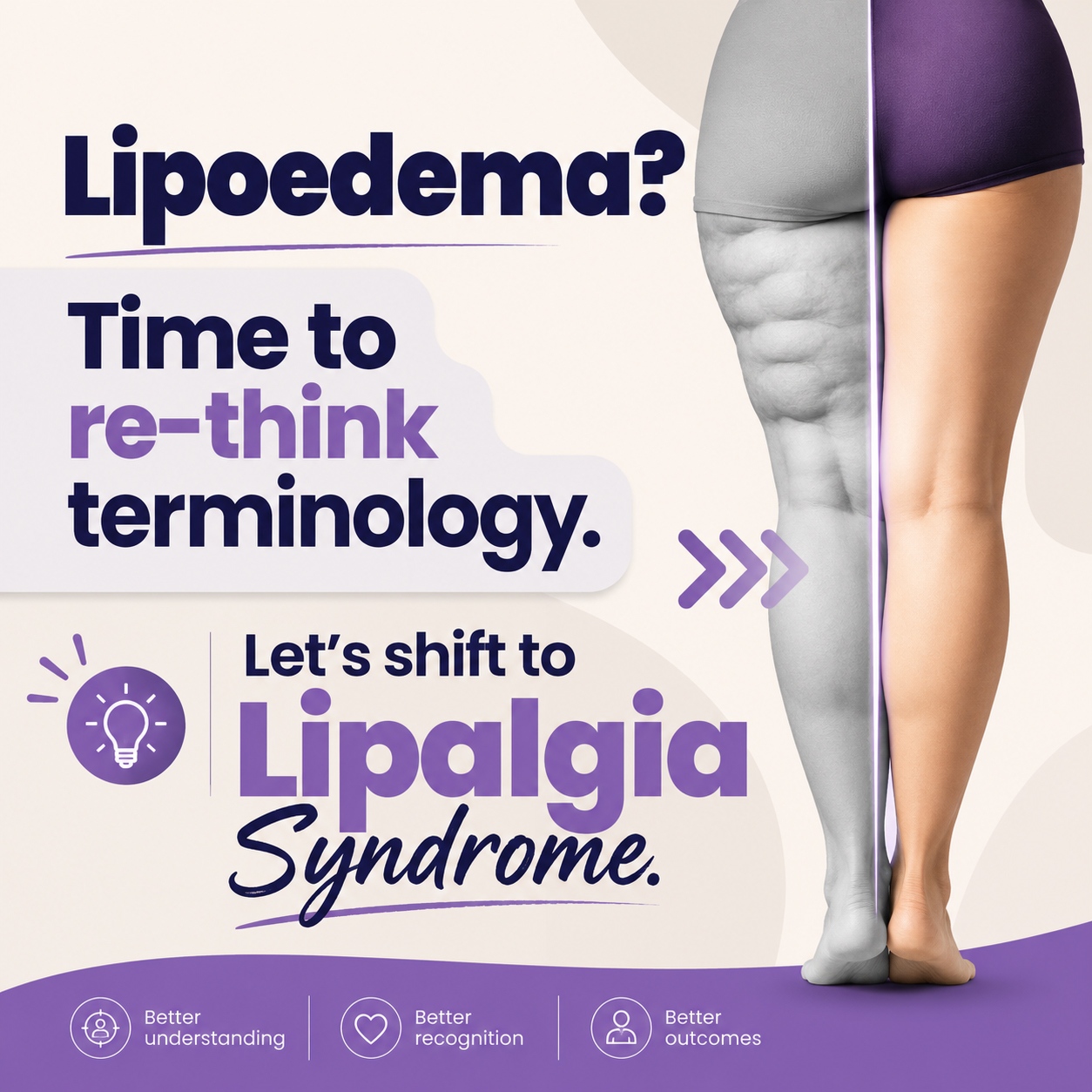
A Scap-Off Load is a special test or functional test that we use to evaluate the implication of Lev Scapular and Up. Trap in Cercival Rotation. As previously mentioned, in cervical rotation, we got a fair bit of muscle working towards this action. As many clients come in with cervical pain, it’s time to explain in more detail what’s going on there. Cervical Rotation. How does it happen? So, when we rotate our head, either right or left, the muscle on the same side of the rotation movement is contracting. If a muscle along those is weak, we may reproduce pain in rotation along the same side. To thin down which muscle is responsible for the limited ROM, we have to safely deactivate some of them to see if the left behind one can deliver the expected movement. Here is an example of how scap offload works. If a client comes in with 30° Cervical rotation on the R and pain on top of the scapula, that could be an indication that its levator scapulae is the muscle to target. To confirm this hypothesis, I would ask the client to shrug their shoulders and flex their elbow (the client is sitting on a stool). After that, I will make my way behind the client, and I will support their shoulder weight with my forearm and hands. As the client relieves the shoulder tension, that lev scapulae and up. Traps. are now deactivated. The next thing would be to ask the client to perform the cervical rotation. Ideally, I would like to see the client have a full range of motion (80° to 90°). If this post talks to you, book your next massage session by clicking here. That would tell me that the only muscles that are limiting the cervical rotation are the lev scap. and up trap. On the other hand, what could happen, is that the cervical rotation is, yes improved, but still limited, compared to the ROM expected. In this case, the muscles involved in the stiff range of motions are not only lev scap. and or upper trap. In fact, what is causing the limitation is the cervical occipital muscles. And yes, spending long hours at the computer or looking at the phone doesn’t help. After this test, to narrow down even more which other muscles are involved in the stiffness of the cervical area, I do run another series of tests. Those tests would look into joint areas like C0-C1, which would refer to Obliquus Capitis Superior muscle, and the C1-C2 test, which would look at tension for Obliquus Capitis Inferior. Furthermore, for the other facet joints that make up the lower cervical region (C3 to C8), I would analyze each facet joint individually. These series of tests are indeed part of my Myotherapy training. Last would be then the usage of the joint mobilisation technique. In this case, we would look into what joint has lost mobility or which one has an excess of it. Strengthening the cervical. In order to improve the presentation, massage on its own is not enough. As per any condition so far, the strengthening of the muscle, in this case, the cervical and upper thoracic one, would allow to prevent further pain and discomfort. The work that the cervical muscle has to do daily is considerably high, giving the natural weight of the skull. So exercising a chin tag in a supine position can help. Ideally, we would do these exercises in the supine position (lying down face up) so that we have gravity to fight back as we train our deep flexors. To further improve the strengthening, once the chin tag is not enough, we can start using a soft rubber band to create resistance. Said so, be mindful that the cervical area is a delicate area to work on too, and those exercises are best practice under the supervision of an expert trainer or massage therapist.
Range of Motion is the movement of a joint within a 3-dimensional space. For each joint, we expect a minimum and a maximum degree of movement. When to use a range of motion evaluation. Before performing a Myotherapy, Remedial Massage, or a Thai massage session, we check for a Range of Motion, also know as ROM. Checking for ROM is to establish the functionality of the joint and the muscles that surround it. For example, when a client walks in complaining of cervical pain, the first thing we look in too after the postural assessment is the ROM. A postural assessment is an evaluation of the skeletal structure. After that, we ask the client to do specific movements with their head. Like, Rotation side to side, flexion, extension, and lateral flexion. Indeed, these are the basic range of motion for the cervical area. What can this range of motion tell us? Well, depending on the essential mobility of the person, we expect a minimum and maximum range. Let’s say that the client has average mobility; we expect the range of motion of their cervical to be: Flexion (able to flex the head forward and leave a gap of 3cm between the chin and the sternum) Extension, we look in too 70° of movement Lateral Rotation we look in too 80° of rotation (the chin is nearly in line with the shoulder) Lateral Flexion we look in too 45°. If this post talks to you, book your next massage session by clicking here. Is the range of motion the same for everyone? A person with a hypermobile joint range may have a 10° about less ROM than this. Indeed, a person that has a hypermobile joint range can reach 10° furthermore. That’s why every person needs his evaluation. Moving forward, if the range of motion is limited, it could be a muscle tightness or a joint mobilization issue. The best approach for improving joint mobilisation is the Myotherapy treatment or Thai Massage. Both techniques relieve muscle tension and improve muscle tone and joint mobility. The release of tension from the muscle would improve the ROM. On the other hand, when we ask a client to do a ROM, we don’t look only for the length of movement. Quality of movement. For the quality of movement, we refer to how smooth the movement is. Is the client trying to compensate for the cervical rotation by flexing the head? Is the client compensating for the flexion by shrugging the shoulder? Indeed, the movement of a joint is the key to understanding what muscle is responsible for the pain, discomfort or limited ROM. Once we individualise the key muscle/s, we can address the issue. Different types of ROM. In conclusion, the ROM can be active, passive and resisted. The client itself does active ROM. Passive are ROM done by the therapist with no assistance in controlling the movement by the client. Reisted is ROM active done by the client, with a resistance force applied by the therapist again the client’s movement. Each of this ROM can tell us something different about the joint. Active is about muscle lengthening. Passive is about joint mobility (ligament and tendon) Resisted is about the strength of the muscle. We can’t use the Resisted if Active or Passive reproduces pain. That would not be safe. Regarding the Resisted ROM, the resistance is applied in 3 different levels. When the pain gets reproduced, we stop the test. Occasionally, an orthopaedic test can be performed too. Orthopaedic tests are specific tests to evaluate in specific the muscle involved in the limited ROM.
Rotator Cuff Muscles and Bursitis. What muscles are the rotator cuff muscles? And why are so important? What happens when the rotator cuff muscles are out of balance? What is Shoulder Bursitis? Firstly, we look in too which 4 muscles are the rotator cuff muscles. Subscapularis Origin Subscapular fossa of scapula. Insertion Lesser tubercle of humerus. Action Arm internal rotation; Stabilizes humeral head in the glenoid cavity. Infraspinatus Origin Infraspinous fossa of the scapula. Insertion Greater tubercle of the humerus. Action Arm external rotation; Stabilize the humeral head in the glenoid cavity. Teres Minor Origin The inferior lateral border of the scapula. Insertion Greater Tubercle of Humerus. Action Arm external rotation, arm adduction; Stabilizes humeral head in the glenoid cavity. Supraspinatus Origin Supraspinous fossa of scapula. Insertion Greater tubercle of the humerus. Action Arm abduction; Stabilization of the humeral head in the glenoid cavity. Now, that we are more aware of the rotator cuff muscles’ anatomy, we can look into their functionalities. So, the rotator cuff muscles’ functionality is to hold the humerus bond in place in the glenoid cavity. Furthermore, if it was not for those groups of muscles when our arm goes into abduction, the humorous head would pop out of the shoulder joint. Indeed, for abduction, we refer to the arm movement, where the arm goes away from the body laterally. As listed above, all those muscles originate from different areas of the scapula. As per result, muscle as Infraspinatus and Subscapolaris are responsible for balancing the scapula along the sagittal plane. For instance, if the Infraspinatus is overtaking in force the Subscapularis, the scapula would result in a winged position. Consequently, this would affect other muscles that insert onto the scapula, for example, Rhomboids, Lat Dorsi etc… Regarding the injuries, as I already mentioned in the blog post “functional test”, the rotator cuff muscles can easily be injured. This is due to the acromion clavicular joint anatomy. Indeed the space between the humeral head and the acromion is quite narrow and hosts what we call Bursa. If this post is talking to you, and you are in need of a massage, book your next session by clicking here. So, a Bursa is a soft bag, that seats between the bonds, allowing tendons to run through the joint without being exposed to pinch between bonds, and keep the bonds separated, avoiding frictions. As per result, by putting the Bursa under repetitive stress, it tends to swallow and get inflamed. Consequently, the tendons that run below the bursa can get squeezed, creating shoulder bursitis, or shoulder impingement.
Functional test and the empty can test. What is a functional test? What is an empty can test and how does it work? Firstly, functional tests are used to test the strength or load capacity of a single muscle. Secondly, the importance of a functional test is due to avoid misinterpretation of the muscle status and joint health conditions. Furthermore, functional tests can be positive or negative. So, for positive, we refer to a test that gave us the result we were suspicious of. For example, if I do an empty can test, and the client during the test complains of pain in the shoulder acromion, the test is positive. But if for instance, the client complains of pain in another area of the shoulder or arm, the test is negative. Even so, as a therapist, we are aware that other area of the arm or shoulder needs to be looked after. What is an “empty can test”? An “empty can test” is a functional test used to validate the state of health of the supraspinatus tendon, at the high of the acromioclavicular joint. In addition, to better understand how this specific test works, let’s look in too the anatomy of the Supraspinatus m. Origin: Supraspinatus fossa of scapula Insertion: Greater tubercle of the humerus Action: Abduct the shoulder and stabilise the humeral head in the glenoid cavity. As per result, the action of the supraspinatus is to laterally elevate the arm and hold in place the humeral head (the Humerus is the bond of the upper arm). Furthermore, the supraspinatus is one of the rotator cuff muscles. The rotator cuff muscles are: Supraspinatus, Teres minor, Infraspinatus and subscapularis. But let’s get back to the empty can test. The empty can test can be done from seated or standing. In addition, the test is conducted in 2 different stages. Initially, we will ask the client to bring the arm in flexion at about 45° and in abduction at 45°. The arm now is sitting aside from the client’s body, on a diagonal line. Now will ask the client to rotate the arm on itself, as if they are emptying a can. As per the result, if at this stage of the functional tests, the client feels pain in the shoulder at the acromioclavicular joint, the test is positive. If that’s not the case, then we can proceed with the resistant part. If this post is talking to you, and you are in need of a massage, book your next session by clicking here. The resistant part consists of placing our hand on the client’s forearm and asking the client to meet the resistance, at 3 different stages. For each stage, the resistance increases and lasts from 3 to 5 seconds. If during any of the 3 stages the client feels pain, at the high of the acromioclavicular joint, the test is positive. But why the client can feel pain during this type of functional test? To answer this question, we have to look in too the acromioclavicular joint anatomy, but I will talk about this topic in the next blog post.


