
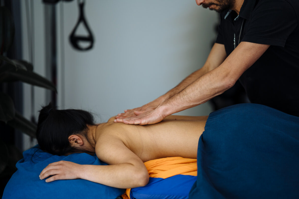
Fascia. What is Fascia? Why is it so important? How does it work?How can Thai Massage help improve fascia mobility? Fascia (from Latin: “band”) is a band or sheet of connective tissue, mainly made of collagen, that seats below the skin and attaches to, stabilizes, encloses, and separates muscles and other internal organs. Firstly, fascia can be classified by layers: Superficial; Deep; Visceral or parietal or by its function and anatomical location. Like as per other body parts, such as ligaments, aponeurosis, and tendons fascia is made from fibrous connective tissues. In addition, these connective tissues contain bundles of collagen fibres oriented in webby patterns, parallel to the direction of pull. About collagen fibres, they are produced from fibroblast, contained within the fascia. In conclusion, fascia differs from other body components such as ligaments and tendons. This is because ligaments hold bonds together and the tendon attaches the muscle to the bonds. Going ahead we can define F. based on its location and Superficial F. This is found just below the last skin layer, subcutis, in most regions of the body, blending with the reticular layer of the dermis. Muscular F. Defined as the fibrous layer seating around the muscle, or other body parts such as nerves, bonds. Visceral or parietal F. This type of structure suspends the organs with their cavities and wraps them in layers of connective tissue membranes. In regards to the functionality of the F., we look in too the ability of this tissue to allow the muscle to move freely. If this post is talking to you, and you are in need of a massage, book your next session by clicking here. So let’s imagine this really thick web, seating between the skin and the muscle and around the muscle too. If the web is overconnected, that means is tight, the muscle would find difficulties in moving and sliding. The same would happen for tendons and organs that need to function by being able to move freely. For example, it does often happen to see customers that when asked to flex the shoulder, as they bring the arm over the head, past the 90° point, it is noticeable a dragging in the fascia along the anterior and or posterior and or lateral thoracic area. The person itself, unless get told to feel what happens in the thoracic area, would hardly be aware of any restrictions along the thoracic area. Said so, we can now open a pandora Jar and talk about the fascia lines. Fascia lines connect the body side to side in the long, for the horizontal and diagonal directions. As per the example, we have an F line that travels from the foot to the head. Indeed this F line starts from the Fibularis M, going up for the IT Band, in too the TFL, Obliquos Abdominal muscles, opposite Serratus Anterior, Rhomboids, and Finally to Splenius Capitis. As per result, someone that suffers from flat feet, and so has Fibularis M, under constant pulling force, may experience shoulder pain on the same side of the body because of Fascia restrictions. But the are more F. lines than the diagonal ones. In fact, we got a Superficial F. line, Functional F. line and Lateral Lines. So, lastly, how Thai Massage can help? Thai Massage can help with stretching the fascia by applying a force on the skin in the direction where the restriction is. Is enough to check in which direction the skin feels tight, and that’s the direction in too we want to apply a stroke with a pulling force, strong enough that the client does actually feel a stretch. At Melbourne Thai Treatment I use techniques of work like MFTT and or Cupping and static/dynamic stretching. In regards to cupping add MFTT are a technique of work that I did learn at RMIT along with my study for the Diploma in Remedial Massage. The static/dynamic stretch, indeed, is part of the techniques used along with the Thai Massage. In conclusion to this blog post, I then would like to invite you to see this video from Dr Gil Hedley, that dives in too the magnificence of Fascia.
Monthly Archives: October 2021

Blog
Simple thing you can do for your Lymphatic System Well-Being. Go For A Walk!
As a therapist who offers Manual Lymphatic Drainage in Melbourne, I am blown away by [...]
Continue readingMay

Blog
Cosmetic Procedure Recovery Melbourne: Liposuction Types and the Benefits of MLD in Fitzroy North
Cosmetic procedures like liposuction are getting more and more popular, with people looking to reshape [...]
Continue readingMay
Blog
Why MLD Before Cosmetic Surgery Matters | Melbourne Massage and Treatment
MLD for cosmetic surgery is often seen as a treatment post-surgery. And while it is [...]
Continue readingMay
Blog
How And Why You Should Look Into Tendon Training?
While training muscles is the most common thing you would see and do in a [...]
Continue readingMay
Blog
Why Your Grip Strength Might Be Predicting Your Lifespan (Yes, Really)
In this blog, we are going to look into why grip strength is one of [...]
Continue readingApr
Oct
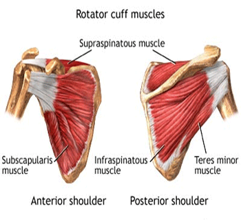
Rotator Cuff Muscles and Bursitis. What muscles are the rotator cuff muscles? And why are so important? What happens when the rotator cuff muscles are out of balance? What is Shoulder Bursitis? Firstly, we look in too which 4 muscles are the rotator cuff muscles. Subscapularis Origin Subscapular fossa of scapula. Insertion Lesser tubercle of humerus. Action Arm internal rotation; Stabilizes humeral head in the glenoid cavity. Infraspinatus Origin Infraspinous fossa of the scapula. Insertion Greater tubercle of the humerus. Action Arm external rotation; Stabilize the humeral head in the glenoid cavity. Teres Minor Origin The inferior lateral border of the scapula. Insertion Greater Tubercle of Humerus. Action Arm external rotation, arm adduction; Stabilizes humeral head in the glenoid cavity. Supraspinatus Origin Supraspinous fossa of scapula. Insertion Greater tubercle of the humerus. Action Arm abduction; Stabilization of the humeral head in the glenoid cavity. Now, that we are more aware of the rotator cuff muscles’ anatomy, we can look into their functionalities. So, the rotator cuff muscles’ functionality is to hold the humerus bond in place in the glenoid cavity. Furthermore, if it was not for those groups of muscles when our arm goes into abduction, the humorous head would pop out of the shoulder joint. Indeed, for abduction, we refer to the arm movement, where the arm goes away from the body laterally. As listed above, all those muscles originate from different areas of the scapula. As per result, muscle as Infraspinatus and Subscapolaris are responsible for balancing the scapula along the sagittal plane. For instance, if the Infraspinatus is overtaking in force the Subscapularis, the scapula would result in a winged position. Consequently, this would affect other muscles that insert onto the scapula, for example, Rhomboids, Lat Dorsi etc… Regarding the injuries, as I already mentioned in the blog post “functional test”, the rotator cuff muscles can easily be injured. This is due to the acromion clavicular joint anatomy. Indeed the space between the humeral head and the acromion is quite narrow and hosts what we call Bursa. If this post is talking to you, and you are in need of a massage, book your next session by clicking here. So, a Bursa is a soft bag, that seats between the bonds, allowing tendons to run through the joint without being exposed to pinch between bonds, and keep the bonds separated, avoiding frictions. As per result, by putting the Bursa under repetitive stress, it tends to swallow and get inflamed. Consequently, the tendons that run below the bursa can get squeezed, creating shoulder bursitis, or shoulder impingement.

Oct
Functional test and the empty can test. What is a functional test? What is an empty can test and how does it work? Firstly, functional tests are used to test the strength or load capacity of a single muscle. Secondly, the importance of a functional test is due to avoid misinterpretation of the muscle status and joint health conditions. Furthermore, functional tests can be positive or negative. So, for positive, we refer to a test that gave us the result we were suspicious of. For example, if I do an empty can test, and the client during the test complains of pain in the shoulder acromion, the test is positive. But if for instance, the client complains of pain in another area of the shoulder or arm, the test is negative. Even so, as a therapist, we are aware that other area of the arm or shoulder needs to be looked after. What is an “empty can test”? An “empty can test” is a functional test used to validate the state of health of the supraspinatus tendon, at the high of the acromioclavicular joint. In addition, to better understand how this specific test works, let’s look in too the anatomy of the Supraspinatus m. Origin: Supraspinatus fossa of scapula Insertion: Greater tubercle of the humerus Action: Abduct the shoulder and stabilise the humeral head in the glenoid cavity. As per result, the action of the supraspinatus is to laterally elevate the arm and hold in place the humeral head (the Humerus is the bond of the upper arm). Furthermore, the supraspinatus is one of the rotator cuff muscles. The rotator cuff muscles are: Supraspinatus, Teres minor, Infraspinatus and subscapularis. But let’s get back to the empty can test. The empty can test can be done from seated or standing. In addition, the test is conducted in 2 different stages. Initially, we will ask the client to bring the arm in flexion at about 45° and in abduction at 45°. The arm now is sitting aside from the client’s body, on a diagonal line. Now will ask the client to rotate the arm on itself, as if they are emptying a can. As per the result, if at this stage of the functional tests, the client feels pain in the shoulder at the acromioclavicular joint, the test is positive. If that’s not the case, then we can proceed with the resistant part. If this post is talking to you, and you are in need of a massage, book your next session by clicking here. The resistant part consists of placing our hand on the client’s forearm and asking the client to meet the resistance, at 3 different stages. For each stage, the resistance increases and lasts from 3 to 5 seconds. If during any of the 3 stages the client feels pain, at the high of the acromioclavicular joint, the test is positive. But why the client can feel pain during this type of functional test? To answer this question, we have to look in too the acromioclavicular joint anatomy, but I will talk about this topic in the next blog post.

Oct
Firstly the Cervical Occipital muscles are a group of muscles, that seat inferior to the skull and are bilateral to the first and second cervical vertebrae. Cervical Occipital muscles are responsible for 45° of rotation out of 90°. Along with these muscles we find: – Obliquus Capitis Inferior; Origin: Spinous process of Axis (C2) Insertion: Transverse process of Atlas (C1) Innervation: Suboccipital nerve – Obliquus Capitis Superior Origin: Transverse process of Atlas (C1) Insertion: Superior line of Occipital bone Innervation: Suboccipital nerve – Rectus Capitis Posterior Major Origin: Spinous process of Axis (C2) Insertion: Inferior line of Occipital bone Innervation: Suboccipital nerve (posterior ramus of spinal nerve C1) – Rectus Capitis Posterior Minor Origin: Spinous process of Atlas Insertion: Inferior Line of Occipital bone Innervation: Suboccipital nerve The actions supported by Occipital Muscles are to extend and rotate the head. How tight Occipital Muscle affect ROM. When rotating the head, so looking at your right or left, you may notice that you don’t have a full range of 80° to 90°, and you start rotating with the thoracic too. One possible reason for this is tension at the occipital area, due to muscle tension and or facet joint irritation. Moreover, to the rotation and extension actions the Cervical Occipital Muscles, are responsible for holding the head up straight. This characteristic is to be taken into consideration with the anti-gravity functionality. In addition to the Cervical Occipital Muscles, other anti-gravitational muscles are the Soleus, Quadriceps Femoris Group, Gluteus Max, and Erectors Spines group. The anti-gravity functionality is essential for the body to fight back gravity (9.81 m/sec2) and allows the body to stand straight up. This group of muscles received information in regard to the gravity pressure from the feet. This information travels along the nervous system starting from the Center of Gravity (COG) of the feet. If the COG is not balanced all body gets affected with a loss of balance as per result. Furthermore, in modern days, the Cervical Occipital Muscles are under constant stress as per all the other anti-gravitas muscles. This is due to spending long hours standing or seating. For example, standing for long hours would overload the feet, leg and back muscles, whereas seating would inhibit the leg muscle but overload the back and cervical muscles. On top of that, spending long hours looking at the phone and or PC would additionally put stress the Cervical Occipital Muscles muscles, as they get over-stretched. How massage can help? As per massaging this area, as therapists, we look into avoiding the Suboccipital triangle, which is defined by the border of the OCI, OCS, and the RCMaj. The Suboccipital triangle is an endangerment site. This means contains superficial, delicate structures that are relatively unprotected and therefore prone to injuries, such as the Vertebral artery, Suboccipital nerve (C1) and Suboccipital venous plexus. If this post is talking to you, and you are in need of a massage, book your next session by clicking here. And what about exercises? As per all the muscles of the body, there are exercises that can be done for the Cervical Occipital Muscles. Along with Thai Yoga, I teach a really simple exercise that recalls the Scap Off Load Test ( a Functional test used to determine what muscle of the cervical region may affect the head rotation). Firstly, in this exercise, available in the Melbourne Massage and Treatment YouTube playlist, you are seating on the floor with a cross leg (a yoga block or pillow can be used as per support), hands projected backwards, with wrists seated below shoulders on a straight line. As per result, the neck would seat in between the shoulders. Secondly, by flexing the head forward, reach the manubrium (the bond that connects the clavicles) with the chin, and with a gentle rotation movement, start rotating the head in a circular movement. Indeed per many Thai Yoga exercises, it is important to be aware of the movement, the body sensation and the speed of movement, which is to be slow and weighted out.

Oct
Remedial Massage and Pichest Thai Massage Technique. Melbourne Massage and Treatment is a Massage clinic that offer services that combines the understanding and approach of Myotherapy with the eastern practice of Thai Massage. I, got into too massage therapy back in 2016, when I did study a Certificate IV in Massage Therapy at the SSNT, in Fitzroy, Melbourne. After that course, I got inspired and directed by a friend to go and study in Chiang Mai, Thailand with Pichest Boonthumme. I could never make a better choice. Was March 2018 when I first started studying with Pichest. But who is Pichest Bonthumme? Why training with Pichest can make a difference? Pichest is probably the most famous and talented Thai Massage teacher/practitioner alive. Pichest was born in Hong-Dong (district south of Chiang Mai) in 1958. He started practising massage in his 20’s at the Chiang Mai Medicinal Hospital, where his father was working as a massage practitioner. It was Pichest’s father who told Pichest himself the basics of Thai Massage. As Pichest was practising and working at the old medicine hospital, he realised that the technique of work that he was using was demanding on his body, till the point he injured his back. From this problem, he creates his strength. He starts working on himself, through meditation practice and devotion to the Buddhist religion, he creates and shaped a new way of massaging and using his body to heal the next one. As you may be aware, Thai people are not so told, and that’s the case of Pichest too. So Pichest massage technique is based on using the body shape and weight to apply pressure on the body of the receiver. Indeed, with this way of working, there is not really a sequence to follow, but there is a pure intuitional approach to go for. Observing the client in their movement and body shape/tension is all you need to start with. Next, come’s the sharpening of the touch. Learn how to feel the tension in the different body parts. In conclusion, once those practices are combined, the Pichest Thai Massage technique is not something that we can achieve in a day or a week of training. We need constant work, in everyday life as it’s more a habit than just a way of massaging, and it’s different from practitioner to practitioner, as we are all different in body size and weight. So where I am with this way of working. I have been studying with Pichest for several months, along with 2018 and 2019. Thanks to previous experience, in the massage industry and elsewhere, I found it easier to start working with the Pichest technique. Said so, there was and still, there are several improvements that I have to achieve. But back to the comparison to a general Thai Massage, Melbourne Thai Treatment offers a more unique way of massaging, as I focus more on the individual needs and not on a general sequence that I can apply to anyone. To reinforce my understanding and knowledge of the body and its way of working I took an Advance Diploma in Myotherapy, at RMIT University. This course allowed me to improve my anatomy knowledge and understanding. If this post is talking to you, and you are in need of a massage, book your next session by clicking here. In addition, I could appreciate even more what I did learn with Pichest, as working on the floor as we do as Thai Massage therapists and working on the table, as Remedial Massage therapists do, are 2 different realities. Working on the floor allows you to move freely and you can use all your body weight as needed to apply pressure. Where, working with a table, you are standing all day long, you must apply force just with your upper body, and the use of hands is demanding, to a point that a massage therapist after few years of practice can injure its hands or wrist badly. Furthermore, working with a Remedial Massage technique style you can use only your hands and elbow to manipulate and massage the receiver. With the Thai Massage, thanks to its intuitional approach you are allowed to use feet, knees, elbows or other body parts. So, the use of knees, feet or other body parts is strictly subjective to the therapist ability and the client needs. It is easier to apply stronger pressure with a knee than a hand probably, but this doesn’t mean that the work done with a knee must be strong all the time. You may wanna use the knee just because you and the client can be in a more ergonomic and comfortable position. But how you can apply the right pressure with the knee? Well, that’s part of our training. Walking with the knee of the wood floor is a start, but also moving from seated to standing on the floor is part of the training. In fact, the Thai Yoga exercises are part of the training that we take as therapists to heal our body from tensions. To finalize, Melbourne Thai Treatment is yes a Thai Massage service, but is based on eastern and western practice and understanding of the body. In order to, improve and enrich my knowledge than, for the upcoming year (2022) I will look into starting a Myotherapist course, always at the RMIT. This is because I would like to add to my practice service as dry-needling and have still a better and more complete understanding of the body and its functionality.
