Strengthening exercises are an important way to improve our overall health by improving muscular endurance and stability around the joints. Unlike isometric exercises, which are performed with static muscle contractions, strengthening exercises involve dynamic movements that are capable of creating contractions and lengthening of muscles. This type of training is relevant not only for sportsmen but also for everyone who wants to raise their physical condition. Why Strengthening Exercises? Recent studies have focused on the role that strengthening exercises play in the maintenance and enhancement of musculoskeletal health. Strength exercises play a key role in preventing injuries, rehabilitation, and the enhancement of daily functional activities. Why this is possible is because this type of training consists of applying resistance to your body that will challenge your muscles, bones, ligaments and tendons, helping build strength, endurance, and overall physical function. Strengthening exercises stimulate muscular hypertrophy, which is defined by an increase in muscle size and strength. This is very important for preventing and even delaying the onset of sarcopenia, a major factor in declining health with natural aging. Strengthening for Injury Prevention and Recovery Imagine having a shoulder injury and undertaking rehabilitation: It would start with light movement and isometric exercises. After the initial healing phase, strengthening integration is essential. Strengthening exercises rebuild the muscle’s strength, provide joint stability, and regain total function. For example, with a shoulder injury, performing resistance exercises such as rotations with a resistance band and or weights, along different planes and directions, can help regain the strength of the rotator cuff muscles. These exercises will enhance not only the muscles around the shoulder but also the overall stability of the joint and consequently reduce the chance of future injuries. Without going through the exercises phase, you may experience a decrease in pain over the weeks after the initial injury (it depends on the severity of the injury), but that doesn’t mean that you are out of danger or re-injury. Indeed, as soon as you place extra force in the joint or on the soft tissue of the shoulder, the risk of re-injury would skyrocket, as the shoulder complex it may not be ready or strong enough to permorm such actions. Body tissues and Exercise Strengthening. What’s the deal? Throughout our lifespan, our metabolism slows down. During this process, not only will we process energy intake differently, but how our body recovers and regenerates will also change. So, all the body’s soft and hard tissues will have difficulty recovering and staying strong. That’s where the strength exercises come through. Applying a resistance to those tissues, a positive stress resistance, would allow those tissues to regenerate and grow stronger. These are valid for both soft and hard body tissues. We define positive stress as something that does not put the body in danger but is a stress that the body can handle and take advantage of, like a few kg of a dumbbell or a resistance band. Then, the stronger we get, the more weight we can handle, and that’s how we progress in the exercises. How Do Strengthening Exercises Benefit Tendons? Probably the most essential rehabilitation of connective tissues besides bones would have to be the tendons, which connect muscles to bones. When you do resistance training, tendons encounter controlled stress that stimulates the building of collagen. Collagen is vital in repairing and strengthening tendons; thus, the substance is one of the priorities in any rehabilitation program for tendons. For instance, in the rehabilitation from Achilles tendinopathy, there are such eccentric calf raises- you gradually lower the heel below the level of the step, which are particularly effective. The exercise improves resilience and strength of the tendon through progressive loading and stimulating collagen synthesis. Examples of Effective Strengthening Exercises Squats: Excellent for overall strength of the lower body. First, try squats with your own weight, then add weights as you progress. Deadlifts: So good for exercising the posterior chain, such as hamstrings, glutes, and lower back. Go with lighter weights to perfect the form before adding any resistance. Push-Ups: A great exercise for your upper body since it targets the chest, shoulders, and triceps very well. Variants such as decline or incline push-ups can be done to increase/decrease the intensity level. Progressing the Strengthening Exercises Exercise progression is necessary if improvement is to be continually achieved; otherwise, this often leads to a level where no significant further improvements are made. Start with exercises that match your current fitness level and gradually increase the intensity by adding more weight, increasing repetitions, or incorporating more complex movements. For example, one can perfect squats with one’s own body weight and then subsequently move on to the next level by adding dumbbells or even a barbell. Or, for instance, when one masters regular push-ups, they can always try modifications, such as adding weights to the push-up or doing one-arm push-ups to make their muscles work harder. Incorporating Strengthening Exercises into Your Routine It is easy to incorporate a routine where another routine already exists. Indeed, strength exercises don’t have to be associated with hours and hours in the gym. You can have a dumbbell or a kettlebell or a resistance band, sitting in your kitchen, and while you wait for your morning coffee to come up, you can do a few squats. A few minutes of well-practised exercise here and there are better than nothing. Start with little, learn how to experience the pleasure of movement and the benefit of exercising and from there you can build a stronger and longer routine of self-care. And as you learn more and more, you can start looking into balancing a comprehensive program that blends strengthening exercises with cardiovascular activities and flexibility training to promote overall health and functionality. How often should we exercise? I often get asked this question when I give exercises to my patients. We now know that the frequency of strength exercises is strictly related to our goals. So to increase your strength, you look for 3 to 5 […]
Tag Archives: massage
Blog
Strengthening Exercises: Building Resilience for Better Health
Strengthening exercises are an important way to improve our overall health by improving muscular endurance [...]
Continue readingBlog
Managing Menopause Symptoms: Lifestyle Changes, Nutrition, Exercise, and More
Menopause is a natural phase in every woman’s life, but it comes with a variety [...]
Continue reading
Blog
Hormones and Menopause: What is happening?
Menopause is a crucial step in a woman’s life. Menopause marks the time when a [...]
Continue readingMar

Blog
How Strength Exercises Can Help With Bone Density
Bone density is a key factor in bond fracture prevention. Bone is made of cells [...]
Continue readingMar

Blog
How Lymphatic Drainage Massage Can Help with Menopause Symptoms?
Menopause is a natural transition in every woman’s life, which marks the end of a [...]
Continue readingMar
Mar
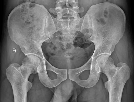
Musculoskeletal pain can be complex, and orthopedic tests and hands-on treatment, sometimes, can be a limited tool to individualise what is happening with the body’s internal structure. Indeed, there are times when a deeper look is required to ensure we are on the right path. This is where body scans imaging comes into play to identify presentations like tendinopathy, bursitis, ligament tear or other underlying conditions. The Role of Body’s Scan in Diagnosing Pathology Body scans include a series of imaging technologies, such as ultrasound, x-ray, MRI, to name a few. Ultrasound is a highly effective imaging tool used to assess soft tissue structures in real-time. Unlike X-rays, which primarily show bone, ultrasound provides detailed images of muscles, tendons, bursae, and ligaments. This makes it an excellent tool for diagnosing conditions such as: Tendinopathy – A chronic condition involving tendon degeneration due to overuse or injury. Bursitis – Inflammation of the bursae, the small fluid-filled sacs that reduce friction between tissues. Those tissue types are found along different body joints, like the shoulder and the hip. Ligament Tears – Partial or complete tears of ligaments, often occurring after trauma or excessive stress. Baker’s cyst – is a fluid-filled swelling that forms behind the knee, often resulting from knee joint conditions like arthritis or meniscal tears, causing discomfort and limited mobility. When we are suspicious of one of those presentations, due to positive results obtained by orthopedic test and medical history, including mechanism of injury, we attempt a recovery process, based on the type of injury, symptoms, and other relevant information. Along this recovery process, we may start with isometric exercises. If, with the first 6 weeks, and a series of sessions, 3 to 4 sessions with this time frame, we still don’t see a major recovery, then we may want to get extra investigation ongoing via an ultrasound scan, which can clarify the underlying pathology. It allows us to confirm or rule out certain conditions, ensuring that treatment strategies are aligned with the actual tissue damage (if any is present). On the other hand, based always on the individual case, we could also require X-rays, which are often more helpful in diagnosing conditions related to the bones, such as arthritis or fractures, as they provide a clear view of bone structure and joint spaces. MRI is a scan that is used for Brain imaging, and when the investigation needs higher details, like when looking at the spine or a joint that via ultrasound was not giving any sign of issue. Ultrasound is also comparable to MRI, as it is faster, easier to deliver, and has fewer complications. How can myotherapy treatment help recovery from what a body scans would show? As we already discussed in another blog, Myotherapy is a practice that looks into the well-being of the skeletal muscle structure. To understand what can be done about a painful presentation, we would initially take a detailed clinical history, then look into objective measurements, such as your movement and body presentation. Given the result we can obtain, we would build up a treatment plan which includes: Hands-on Treatment – Techniques such as deep tissue massage, myofascial release, and dry needling can help reduce pain and improve mobility. Exercise Prescription – Strengthening and mobility exercises help restore function and prevent future injuries. Load Management Strategies – Proper guidance on activity levels ensures tissues heal without excessive strain. That management technique would then be combined and adjusted around the scan’s results. Here are a few examples: Bursitis: If a bursitis is confirmed, medications may be given to reduce the inflammation of the bursa, for that, we concentrate on MLD treatment to further reduce the inflammation and exercises to build strength on the structure that needs support. Ligament tear: When talking of ligament tear, the healing time can dilagate to months if not also a year, so we know now why the 6 weeks program may was not as responsive. We will keep focusing on the strength of the muscle that surrounds the specific joint, and use hands-on treatment to boost blood to the area affected. Arthritis: Medication or dietary change may be put in consideration for pain management and inflammatory reduction. Also in this case, MLD can be used to manage the pain response, and exercises for mantain movement in the affected joint/s. When Should You Consider an Ultrasound or other body scans? If you experience ongoing pain, swelling, or restricted movement that is not improving with therapy, an ultrasound or other scan helps identify the cause. This can prevent prolonged discomfort and allow for a more targeted treatment approach. At Melbourne Massage and Treatment, in Fitzroy North, we aim to provide the most effective care possible. If you’re dealing with persistent musculoskeletal pain, book a consultation with Giovanni today. Together, we’ll determine the best action to get you back to optimal function. Frequently Asked Questions (FAQs) About Musculoskeletal Pain and Body Scans Imaging 1. What are body scans, and how do they help diagnose musculoskeletal pain?Body scans include imaging technologies such as ultrasound, X-ray, and MRI. These scans help diagnose soft tissue injuries (like tendinopathy, bursitis, and ligament tears) or bone-related conditions (such as fractures or arthritis). They provide a clearer picture of what might be causing pain, inflammation, or restricted movement. 2. Why is ultrasound commonly used in diagnosing soft tissue injuries?Ultrasound is highly effective for real-time imaging of soft tissues like muscles, tendons, bursae, and ligaments. It helps diagnose conditions such as tendinopathy, bursitis, and ligament tears, providing a dynamic view of the area being studied without the need for invasive procedures. 3. When should I consider getting an ultrasound or other scans for my injury?If you’re experiencing persistent pain, swelling, or limited mobility that isn’t improving with initial therapy (such as exercises or hands-on treatment), it might be time to consider an ultrasound or other scans. These can help identify the underlying cause of your symptoms and allow for a more targeted treatment approach. 4. How do orthopedic […]

Mar
Meralgia Paresthetica is a condition characterized by numbness, tingling, and burning pain in the outer thigh. It occurs when the “lateral femoral cutaneous nerve” (LFCN), which supplies sensation to the skin of the thigh, becomes compressed or irritated. While not life-threatening, it can be uncomfortable and disruptive to daily activities. Now let’s see how Myotherapy treatment can help with this presentation. Causes of Meralgia Paresthetica Meralgia Paresthetica presents with symptoms of lateral thigh weakness and numbness, which are caused by compression or the lateral femoral cutaneous nerve. This compression can have typical causes, which include: Tight clothing – Wearing tight jeans, belts, or shapewear can compress the nerve at the hip height. Obesity or weight gain – Excess weight puts pressure on the nerve. Pregnancy – The growing uterus may contribute to nerve compression. Prolonged standing or walking – Excessive movement can irritate the nerve. Injury or trauma – Previous surgeries, injuries, or direct impact to the hip area can damage the nerve. Diabetes – Diabetic neuropathy may increase susceptibility to nerve-related conditions. Inguinal ligament – The LFCN passes right under the inguinal ligament, which may create compression on the nerve itself. All those factors can compress the branch of the femoral nerve, which innervates the lateral portion of the thigh. The origin of the nerve is at the lumbar level L2/L3. Indeed, when a portion of the skin has altered sensation, it is often a peripheral compression that causes the symptoms. Symptoms of Meralgia Paresthetica The symptoms for Meraglia Parestetica often involved one leg only, as it is quiet uncommon to get compression bilaterally. Those symptoms include: Burning, tingling, or numbness in the outer thigh. The skin of the lateral thigh can also become very sensitive and painful to the touch. Sharp or aching pain that worsens with prolonged standing or walking. If the compression is due to organs or the inguinal ligament, movement can aggravate the presentation due to the tightness of the structure during movement. Increased sensitivity to touch in the affected area. Muscle weakness is not a symptom, as this condition affects sensation, not motor function. How can myotherapy treatment help individualise this presentation? As a myotherapist, I specialise in muscular skeletal presentations, and we focus on soft tissues. Through a series of assessments, we can determine whether the compression is peripheral or root nerve compression. Let’s see how. Medical History In the first step, we examine the medical history and physical examination, identifying risk factors and symptoms. Along with the physical examination, we examine the Myotome and Dermatome. Examination On top of active range of motion and another orthopedic test to rule in and out other possible presentations, there are some specific tests which we want to focus on, Myotome and Dermatome. The Myotome are resisted movement, like in this case, hip flexion, knee flexion, knee extension, where it would result in positive findings if we have evident weakness and or back pain. This test would rule in a compression to the spine level. Dermatome, on the other hand, are used to test the connectivity of the cutaneous nerve. So with a sharp and soft object, we will mark some line along the thigh area, looking for loss or confused sensations. This test would rule in a peripheral compression of the nerve. Notice that both presentations can be presented at the same time. Other tests that can be done for this presentation include: Electromyography (EMG) – To rule out other neurological disorders. Imaging tests (MRI, X-ray, or ultrasound) – Identifying structural issues or nerve compression. For those tests, Giovanni would write a referral letter for your GP. Treatment Options for Meralgia Paresthetica As often happens, the treatment options are multiple and must be embraced in groups, not individually. The overall aim of any treatment is to relieve pressure on the nerve and reduce symptoms. Here is a list of treatment options and modalities: Lifestyle Modifications Wear loose-fitting clothing to reduce nerve compression. Weight management to decrease excess pressure on the nerve. Avoid prolonged standing or walking if symptoms worsen. Medical Treatments Pain relievers – NSAIDs (like ibuprofen) or acetaminophen for mild pain relief. Myotherapy treatment – along a series of myotherapy sessions we can reduce symptoms and improve the presentation. Corticosteroid injections – Reduce inflammation and pain. Nerve blocks – In severe cases, numbing the nerve can provide relief. Surgical Options (For Severe Cases) Nerve decompression surgery – Relieves pressure on the nerve. Neurectomy – Removing the affected nerve if pain is persistent. How Myotherapy Can Help At Melbourne Massage and Treatment, during a myotherapy session, after ensuring we are dealing with a Meralgia Paresthetica I may use a series of techniques to help you out with your symptoms. What technique to use is based on your individual presentation,, other cohexsitng presentations, adn also your choice and comfort. Here is a list of modalities used during a Myotherapy session: Muscle Energetic Technique (MET) – Helps reduce tension in the hip, thigh, and lower back muscles that may be contributing to nerve compression. Trigger Point Therapy – Addresses myofascial trigger points that can exacerbate pain and discomfort. Mobility and Strengthening Exercises – Improves mobility and reduces pressure on the nerve. Postural Education – Helps correct movement patterns that may be aggravating symptoms. Joint Mobilization – Enhances circulation and reduces inflammation in affected joints. Dry needling – Using a needle can help reduce pain and muscle ache and increase the neurological connection of those same soft tissues. After the hands-on treatment, we will then look into exercises that can help maintain the change we created. That said, there are other precautions to take in consideration, like: Maintain a healthy weight to prevent excess pressure on the nerve. Choose comfortable clothing that doesn’t constrict the waist or thighs. Incorporate gentle mobility and exercise into your routine. Monitor underlying conditions, such as diabetes, to reduce nerve-related complications. Conclusion Meralgia Paresthetica can be managed effectively with lifestyle changes, medical treatment, and preventive care. Myotherapy can be a valuable complementary […]

Feb
Whiplash is a common neck injury caused by a sudden and forceful back-and-forth motion of the head. We often see this in patients who go through a car accident, contact sports injuries (AFL, rugby or even Soccer), or falls. While whiplash is usually not life-threatening, we now know that it can cause persistent pain and discomfort, affecting daily activities. Therefore, it is important to understand its symptoms, causes, and the best exercises for recovery, which can help individuals manage and overcome this condition effectively. Common Causes of Whiplash Whiplash is a term used to describe a fast rocking motion of the cervical area and is most frequently caused by: Car accidents: Rear-end collisions are the leading cause, as the sudden force propels the head forward and backward. Sports injuries: Contact sports like football, boxing, or hockey but even AFL, Rugby or even Soccer increase the risk of whiplash injuries. Falls: Slipping and falling can cause the head to jerk suddenly, leading to whiplash. Physical assaults: Blows to the head or sudden jolts, such as those experienced in shaken baby syndrome, can result in whiplash. What are the consequences of Whiplash for the cervical ligaments? The consequences of whiplash for the cervical ligaments can be significant, leading to long-term instability and chronic pain. When the ligaments are overstretched or torn, they lose their ability to support the cervical spine, resulting in properly: Reduced Stability: Weakened ligaments can no longer provide adequate support to the cervical vertebrae, leading to excessive movement and an increased risk of further injury. That’s why stretching is not recommended either. Chronic Pain and Stiffness: Persistent discomfort may arise as the muscles attempt to compensate for the lack of ligament support. Increased Risk of Degeneration: Ligament damage can accelerate wear and tear on the cervical joints, potentially leading to conditions such as osteoarthritis. Neurological Symptoms: Instability in the cervical spine may irritate or compress nerves, leading to headaches, dizziness, or numbness in the arms. Symptoms of Whiplash Whiplash symptoms can range from mild discomfort to severe pain, and they often appear within hours or days of the injury. Common symptoms include: Neck pain and stiffness: One of the most prevalent symptoms, often worsening with movement. Headaches: Typically originating from the base of the skull and radiating toward the forehead. Shoulder and upper back pain: The impact can cause muscle strain in surrounding areas. Reduced range of motion: Difficulty moving the neck due to stiffness and discomfort. Dizziness and fatigue: A common reaction as the body copes with the injury. Tingling or numbness in the arms: Nerve involvement may lead to sensations of pins and needles. Cognitive issues: Some people experience memory problems, difficulty concentrating, and irritability. Those symptoms may not present all at once, and they can belong to other presentations, while whiplash did or didn’t happen anytime before. That’s why when we go through a clinical history taking, as Myotherapist, we take our time to dig into your past and your body habits, as this can give us important information about your current presentation and what we can do to improve it. Why Can Whiplash Become a Lifelong Issue? In some cases, whiplash can become a chronic condition due to the instability of cervical ligaments. Cervical vertebrae can be divided into two parts, Mobile and Stable joints. C1 to C2 are the mobile ones, whereas C3 to C7 are the stable ones. Now, if along a whiplash incident, the ligaments of either the mobile or, most luckily, the stable side get strained, the muscles surrounding that segment of the joint would have to work harder to maintain stability. This would lead to to ongoing discomfort, reduced mobility due to pain and muscle spasms, and increased vulnerability to future injuries. If left untreated or managed improperly, this is how the condition can become chronic and lead to other injuries along the way. Importance of Thoracic Mobility Thoracic mobility plays a crucial role in preventing the chain effect of mobility and stability issues between the thoracic spine, lower cervical, and upper cervical regions. If the thoracic spine is stiff or restricted, the lower and upper cervical spine must compensate, leading to increased strain and pain. Improving thoracic mobility through targeted exercises can help reduce this compensatory stress, allowing for better neck function and reducing the risk of chronic discomfort. In a case of a previous history of whiplash, maintaining good thoracic mobility would allow the cervical area to focus on its duties, so stability for the lower portion and mobility for the upper. Reducing the risk of overcompensation and muscle fatigue. Effective Exercises for Whiplash Recovery A structured exercise program is essential for whiplash recovery. This program should begin with isometric exercises to restore basic cervical movement, progress to concentric exercises to rebuild strength, and eventually include thoracic mobility drills to enhance overall spinal function. Phase 1: Isometric Exercises for Early Recovery. For the first 2 weeks post-injury, 5 to 7 days a week. Isometric exercises help activate muscles without excessive movement, providing a stable foundation for recovery. This step is essential to start driving more blood to the area irritated by the whiplash and also allow the central nervous system to feel confident in perceiving the cervical structure moving without pain. Isometric Neck Holds: Place your hand on your forehead and gently press against it without moving your head. Push only 25% of your strength, as it has to be a pain-free exercise. Hold for 5-10 seconds and repeat 5 times. These exercises can be done in any cervical movement, such as extension, lateral flexion, or rotation, by using your hand as a resistance and pushing always at 25% of your strength. The strenght of push does not ever progress, what you will progress within isometric exercises are the time fram of push, repetitions and sets. Phase 2: Concentric Strengthening Exercises. From week 3 post-injury onwards,3 to 5 times a week. Once the pain subsides, which we would expect to happen in 2 weeks about it, gradual […]

Feb
Greater Trochanteric Pain Syndrome (GTPS) is a common condition that causes persistent lateral hip pain, often making everyday activities like walking, climbing stairs, or even lying on your side difficult. GTPS primarily affects middle-aged individuals, particularly women, and is commonly linked to issues such as gluteal tendinopathy and weakness in the hip stabilizing muscles. At Melbourne Massage and Treatment, our focus is on evidence-based approaches to managing GTPS, and the latest research strongly supports the role of exercise as the first line of treatment for this condition. GTPS Symptoms Greater Trochanteric Pain Syndrome can present with a series of symptoms that are local to the side of the hip. Here are the most common: Lateral hip pain: Persistent pain on the outer side of the hip, which may extend down the thigh. Pain when lying on the affected side: Discomfort that worsens when lying directly on the hip. Tenderness to touch: Sensitivity around the greater trochanter, which may be painful to press. Pain with movement: Aggravation of pain during walking, climbing stairs, or standing for prolonged periods. Weakness in hip muscles: Reduced strength in the gluteal muscles, leading to instability in movement. Difficulty sitting for long periods: Sitting on hard surfaces can exacerbate discomfort. Mechanism of Injury for GTPS GTPS is primarily associated with tendinopathy of the gluteus medius and/or minimus muscles, with or without accompanying bursitis. As per many tendon injuries, this condition often arises from repetitive stress or overuse, leading to microtrauma and degeneration of these tendons. On the other hand, abnormal hip biomechanics can exacerbate the issue, as compressive forces cause impingement of the gluteal tendons and bursa onto the greater trochanter by the iliotibial band during hip adduction. Contributing factors to GTPS include acute trauma, such as a fall onto the lateral hip, prolonged pressure from lying on one side, and overuse from activities like running or stair climbing. Additionally, conditions like iliotibial band disorders and gluteal muscle weakness can increase the risk of developing GTPS. Understanding these mechanisms is crucial for effective management and prevention of GTPS. Evaluation of GTPS Diagnosing GTPS typically involves a combination of clinical examination and medical history assessment. After taking your clinical history, including sports and work activity, I will perform a series of tests to validate the suspicions of GTPS. Those tests include single-leg stance and resisted hip abduction, which we would expect to show weakness in single-leg standing and pain during the abduction movement. Lastly, we would also palpate the area, which is a test that is kept for last because we want to avoid flair the presentation, which may be painful with any other test after that. In some cases, imaging techniques like ultrasound or MRI may be used to rule out other conditions and confirm gluteal tendinopathy or soft tissue abnormalities. I personally do not recommend image testing as the first way to go because the impact of seeing physical damage can also have a negative impact on self-perception, making a recovery harder. At Melbourne Massage and Treatment, our focus is on evidence-based approaches to managing GTPS, and the latest research strongly supports the role of exercise as the first line of treatment for this condition. The difference between GTPS and Femoroacetabular Impingement (FAI) The difference between GTPS and FAI stands in the hip area involved in the injury. The GTPS is relative to the side of the hip and involves the gluteus medius and minimus tendon and the bursa that separate that tendon from the greater trochanter of the femur. On the other hand, FAI is a presentation that still involves the hip, but it does take place on the anterior portion of the hip, as is characterised by and overgrowth of tissue on the femur head or the hip socket, and it does manifest with hip flexion and external rotation. That’s why it is important to receive an evaluation of the presentation from a professional, in order not to mix the two presentation, or also, in order to evaluate if both presentation are present at the same time, which can also happen. The Role of Exercise in GTPS Treatment A recent systematic review and meta-analysis analyzing multiple randomized controlled trials found that structured exercise provides significant benefits for individuals with GTPS. The findings revealed that: Long-term pain reduction: Exercise can lead to slight but meaningful reductions in hip pain over time. Improved physical function: Patients who engage in targeted exercise programs experience better mobility and overall hip function. Increased likelihood of meaningful recovery: Compared to corticosteroid injections, exercise significantly increases the chances of noticeable improvement in symptoms. One of the most notable takeaways from this research is that exercise has a long-lasting effect, whereas treatments such as corticosteroid injections may provide only short-term relief. Additionally, no serious adverse effects were reported with exercise-based interventions, making it a safe and sustainable approach to managing GTPS. Why Choose Exercise Over Corticosteroid Injections? Corticosteroid injections have often been used for GTPS pain relief, but the research indicates that exercise leads to better long-term outcomes. While injections may offer temporary symptom relief, they do not address the underlying causes of GTPS, such as gluteal muscle weakness or tendon dysfunction. Exercise, on the other hand, strengthens the hip muscles, improves joint stability, and reduces the likelihood of recurring pain. In a previous blog post, I spoke about the key role of Gluteus Medius as a pelvis stabiliser. Effective Exercises for GTPS At Melbourne Massage and Treatment in Fitzroy North clinic, I design individualized exercise programs to help patients with GTPS regain strength and function. Some of the most effective exercises for GTPS include: Isometric exercises: Holding static positions to engage the hip muscles without excessive movement, reducing pain and improving muscle endurance. Strength training: Progressive strengthening of the gluteus medius and minimus muscles to enhance hip stability. Functional movement training: Exercises that mimic daily activities to help improve movement patterns and prevent pain triggers. These exercises can be performed both at home and under professional supervision to ensure […]

Feb
When it comes to maintaining a healthy, functional body, it’s easy to overlook the pivotal role of certain muscles in everyday movement and long-term stability. One such muscle is the gluteus medius. At Melbourne Massage and Treatment, located in Fitzroy North, I see many patients who either love running or love to hit the gym but are not aware of the importance of this muscle for their activity. What is the Gluteus Medius? The gluteus medius (GM) is one of the three primary muscles of the gluteal group, located in the upper part of the buttock. Here is a breakdown of its anatomy: Origin: the gluteal surface of the ilium Insertion: lateral surface of the greater trochanter Innervation: dorsal branches of the L4, L5, and S1 Actions: Abduction and medial rotation of the lower limb. It stabilises the pelvis. Thanks to its positioning, the GM plays a vital role in controlling pelvic movement, specifically in the stabilization of the pelvis during various motions like walking, running, or standing on one leg. More Information About Gluteus Medius actions The GM serves several essential functions that directly affect the stability of the hip and lower body: Pelvic Stabilization: One of its primary roles is preventing the pelvis from tilting excessively to one side when you move, especially when you’re walking or running. If the gluteus medius isn’t working properly, the opposite side of your pelvis may dip downward, leading to an imbalance and compensatory movements that strain other parts of the body. Hip Abduction: The gluteus medius helps to move the leg out to the side, away from the body. This movement, known as hip abduction, is crucial for activities that require lateral movement, such as stepping sideways or maintaining balance while performing physical tasks. Internal and External Rotation: The gluteus medius also assists with the rotation of the hip joint. Depending on which fibers are activated, it helps with both internal and external rotation of the thigh. This is essential for maintaining control and precision in movements. Postural Support: The gluteus medius muscle helps keep the pelvis level when you’re standing on one leg. Without proper activation of this muscle, one hip might drop, affecting posture and causing misalignments in the spine and lower back. The Role of the Gluteus Medius in Hip Stability Why is the GM so important for hip stability? Simply put, this muscle acts as the stabilizer of the pelvis. Without a properly functioning gluteus medius, other muscles and joints are forced to compensate for the lack of stability, leading to overuse and strain. For example, improper GM function can result in excessive stress on the knees, lower back, and even the ankles, which can lead to pain, discomfort, and injury. Clinical implications are vast, especially for athletes and individuals who regularly engage in physical activities. Hip instability can result in difficulty performing simple tasks like walking or climbing stairs, and over time, it may contribute to chronic conditions such as hip osteoarthritis. A common painful presentation that we see in athletes but also the everyday patients is Greater Throcanta Pain Syndrome (GTPS), which is characterised by the side hip pain. This presentation results from a GM tendon irritation. Signs of Weak or Dysfunctional Gluteus Medius Here are some common signs that your gluteus medius may need attention: Pain in the hip or lower back: Since this muscle is integral to proper alignment, dysfunction often manifests as discomfort in the hips or lower back. Difficulty balancing on one leg: Struggling with stability when standing on one leg may indicate weak gluteus medius muscles. Shifting or limping while walking: A noticeable shift or limp while walking can point to weakness in the gluteus medius, causing the body to compensate and disrupt your gait. How can Gluteus Medius impact your run? The gluteus medius is crucial for runners as it stabilizes the pelvis, controls hip movement, and ensures proper alignment during running. This muscle prevents excessive pelvic tilting, reduces side-to-side sway, and helps maintain efficient running form, thereby lowering the risk of injuries such as knee pain, IT band syndrome, and lower back discomfort. A weak or dysfunctional gluteus medius can lead to compensatory movements, affecting performance and causing imbalances. How Melbourne Massage and Treatment Can Help At Melbourne Massage and Treatment, as a clinical myotherapist, I focus on treatment designed to address muscle pain and dysfunction through a variety of techniques. One of the key areas of focus is to create a treatment plan that works for your presentation based on your clinical history. Here is a breakdown: 1. Assessment and Diagnosis: I would conducts a thorough assessment to identify if the gluteus medius is underperforming, weak, or compensating due to other musculoskeletal issues. This involves a combination of posture analysis, movement patterns, and targeted strength tests. 2. Myotherapy Treatment Techniques: I will use various techniques, including trigger point therapy, Dry Needling, myofascial release, and deep tissue massage, to release tension in the gluteus medius and surrounding muscles. This helps to restore proper function, reduce pain, and improve mobility. 3. Rehabilitation and Strengthening: After addressing any issues, we will work to develop rehabilitation strategies, including targeted strengthening exercises for the gluteus medius and other muscles that surround the pelvic, lower back and leg area. These exercises aim to restore proper muscle activation and prevent future imbalances. 4. Injury Prevention: In order to prevent further injury, we will set a target of strength that you want to achieve with your sports activity, and we will do our best to hit that target. Be mindful that based on your presentation, the target could extend from a few weeks to several months. The Takeaway The gluteus medius muscle is far more important for hip stability than many people realize. Its role in maintaining pelvic alignment and controlling movement is essential for pain-free mobility, proper posture, and long-term musculoskeletal health. Whether you’re dealing with hip pain, experiencing difficulty with balance, or want to prevent future issues, understanding and caring for […]

Jan
A deadlift (conventional deadlift) is a popular exercise that aims to strengthen your posterior chain muscle, including the erector spinae muscle, glutes, and hamstring. It is considered a really top list of important exercises to do, and it can be fun and rewarding, but when the weight you move starts increasing, it can lead to severe injuries if you are not using the right technique. Let’s then look into what we need to do to get a good deadlift by starting to analyse from bottom to top how the body should be placed. Centre of mass and biomechanics in deadlift To start with, let’s talk about the biomechanics and the centre of mass for a deadlift. When doing exercises, biomechanics plays a crucial role in safety and optimal exercise execution, and there is no exception for the deadlift. Furthermore, along with all exercises, the lifting and the descending part, the weight has to be in line with the centre of mass. These two components are strictly interconnected to the other one, which means if I don’t use the right biomechanics, I am not going to have the weight aligned with the centre of mass, or if the weight is not aligned with the centre of mass, I am not using my biomechanics at its full potential. So, what’s the centre of mass in the deadlift? The centre of mass in a deadlift is that imaginary line that runs right from the mid-portion of your feet up right in front of your shins, and as you lift the weight up, it passes right in front of your pelvis. That’s where the bar is going to end once you complete the lifting motion. What happens if I don’t keep the bar along the centre of mass? At any stage, during the lifting or the descending motion, if you move the bar further away from the centre of the mass line, there is a great danger of injury. This is because, as the weight travels away from the centre of mass, there is an increase in momentum, which means that your muscles and ligaments that are working hard to move the weight are suddenly placed under a greater load. What are then the proper biomechanics to observe along a deadlift? Ankle and Knee To execute a good deadlift, we want to ensure we have good ankle dorsiflexion, which is not as important as when we squat, but still, we better ensure it is working right. This would allow a straight forward movement of the knee, which would not need to find its way medially or laterally along the initial bending for when we go to grab the bar. Moving up the chain, as we said, the knee have to point straight ahead, following the toes direction. Hip and lower back Next is the hip. This is an important joint, and here is where we need to make sure that we tilt the pelvis forward (bring the teil bone upwards) and as we hinge the hip, we have to have enough movement in there that the greater trochanter (bone landmark that represents the side of the femur’s head) is posterior to the malleolus (the bone landmark that make the side and medial portion of the ankle). Now, if we managed to have tailbone project far back and up, and hip hinged with a slightly bent knee, our back up to cervical area would be alrady quiet flat. Thoracic and head If we keep going upwards, we get to the upper thoracic area, right between the scapula. Here, we want to keep the scapula protracted and have the rhomboids and serratus anterior muscles active and strong so that the arms can hang down straight towards the bar and sit right next to the knee. Regarding the head, use your eyesight to look down at the floor at 45° in front of you and feel the ears pulling away from the shoulder. That would keep your neck nice and long and place the head in the right position. Arms and hands Arms hang down from the shoulder in a straight line, from the AC Joint down to the wrist. The arm has to feel heavy and prolonged, and the hands must sit right next to the shin. Indeed, your arms must stay as wide as your shoulders. That would ensure that your arms are at 90° with the bar, and from a vertical pool point of view, they can take the maximum load ever. Lastly, regarding the hands, there are different grip types that can be used for the deadlift. What is most important is that the wrist is straight following the armline. For exercise purposes, you can have a regular grip where your thumb is gripping around the bar. For heavier weights, you may want to do a mixed grip, where one hand (the dominant one) has the palm facing forward, and the other hand has the palm facing you. In conclusion, the biomechanics of the deadlift, if used correctly, will allow you to always weight in a safe spot, in line with the centre of mass. Your back has to be flat at all times, and along the exercise execution, you want to grasp air in, engage the core to flat out the lower back and then you can lift off. Benefits of Deadlifting Now, let’s look into the benefits of deadlifting. Full-body workout: Deadlifts engage multiple muscle groups—glutes, hamstrings, quads, lower back, core, traps, and forearms—providing a full-body workout in a single movement. Improved strength: Deadlifts are among the best exercises for building overall strength, especially in the posterior chain (back, glutes, and hamstrings). Better posture: Deadlifts can improve posture and reduce the risk of slouching by strengthening your back and core muscles. Core stability: The movement requires significant core activation, helping to enhance core strength and stability. Increased athletic performance: Deadlifts translate well to other athletic movements, as they improve explosiveness, agility, and endurance. Fat loss: The intensity and demand on […]

Dec
When the lymphatic system stop working, either because for a congential malfunctioning, or because of a an external intervention, which did lead to lymphatic system damage, the individual may start experiencing symptoms of Lymphoedema. At Melbourne Massage and Treatment in Fitzroy North, Giovanni understand how challenging lymphoedema can be, which is why he provide expert care through Manual Lymphatic Drainage (MLD) and Combined Decongestive Therapy (CDT). These non-invasive treatments help manage the symptoms and improve your quality of life. What is Lymphoedema? Lymphoedema occurs when the lymphatic system is blocked or malfunctioning, leading to a buildup of lymphatic fluid. This fluid results in swelling in the affected areas, often in the arms, legs, or other extremities. Lymphoedema can be primary (a hereditary condition) or secondary (often caused by injury, surgery, or infection). The fluid that builds up in the body is a fluid that is naturally produced by the body and is released under the skin and between tissues by the bloodstream. It is reached in protein, bacteria, viruses, dust, and other minor substances that the body is unsure how to handle. Stages of Lymphoedema Lymphoedema progresses in stages, with each stage representing the severity of the condition. Recognizing the symptoms early on can help prevent the condition from advancing, so it’s important to be aware of the subtle changes in your body. Stage 0: Latent or Subclinical Stage In this early stage, there are no visible signs or symptoms of lymphoedema, but the lymphatic system may already be compromised. People in Stage 0 may experience a feeling of heaviness, discomfort, or mild swelling, int the limbs affected by this presentation, but these symptoms typically disappear after resting or with limb elevation. The fluid retention is still minimal and may not be noticed by the individual, but it can be detected through careful assessment. Stage 1: Reversible Stage At this stage, swelling is more noticeable, but the skin is still soft, and the swelling can still decrease with limb elevation and movement. When the swelling is present, the skin will feel puffy or tight, and there may be a sensation of heaviness in the affected area. In this relevant early stage, it i still easy to intervene for prevent further degeneration, and if you are unsure of what you are experiencing, reach out Giovanni for a 15 minutes free consultation, in which, at least thanks to the analysis of your clinical history, we can already evaluate if what you are experiencing is a Stage 1 Lymphoedema. Stage 2: Spontaneously Irreversible Stage In Stage 2, the swelling becomes more persistent and is not fully responsive to movement and limb elevation. The asking of the affected area may begin to feel firmer, and there can be noticeable thickening of the skin. This is the stage where fibrosis (scarring of the tissue) starts to develop, and it’s crucial to stop this from going any worse. While the swelling might fluctuate, it becomes more difficult to manage without intervention. At this point, manual treatments like Manual Lymphatic Drainage (MLD) can significantly help reduce swelling and improve the overall function of the lymphatic system. Stage 3: Lymphostatic Elephantiasis The final stage of lymphoedema is characterized by extreme swelling and thickened, hardened skin. The affected area may look large, disfigured, and become painful to the touch. Tissue fibrosis is advanced, and the skin may develop ulcerations or infections due to poor circulation and immune system function. Indeed, along with the swelling and the fibrosis buildup, the outer layer of the skin is placed further away from the blood capillary, which is responsible for releasing oxygen and other substances essential for skin regeneration.Therefore, as the skin breaks open, due to its poor condition, it is more subject to infections and contamination of pathogens. Stage 3 lymphoedema is debilitating and requires ongoing care and treatment to prevent complications and manage symptoms. At this stage, a combination of therapies such as Combined Decongestive Therapy (CDT) and MLD becomes vital for managing the swelling and restoring lymphatic flow. Common Symptoms of Lymphoedema Lymphoedema symptoms can vary from person to person and depend on the stage of the condition. The common symptoms include: Swelling: The most obvious sign of lymphoedema is swelling in the affected area, typically starting in the arms or legs. The swelling is often gradual and may worsen over the course of the day, especially after prolonged standing or sitting. Tightness or Heaviness: Affected limbs may feel heavy or tight, particularly after physical activity or at the end of the day. This sensation is often worse in the early stages and may become more pronounced as the condition progresses. Pain or Discomfort: Pain, tenderness, or discomfort in the swollen area is common, especially when there is fibrosis (hardening) of the tissues. The pain can range from mild to severe, depending on the stage. Reduced Range of Motion: As the swelling and fibrosis increase, it can lead to a limited range of motion, particularly in the arms and legs. This can impact daily activities and mobility. Skin Changes: In later stages of lymphoedema, the skin may appear thickened or leathery, with a shiny, tight appearance. There may also be visible folds in the skin, particularly around the knees, elbows, or ankles. Frequent Infections: Swollen tissues have a reduced ability to fight off infections, so people with lymphoedema are more susceptible to bacterial and fungal infections, which can further complicate the condition. Numbness or Tingling: As the swelling progresses, the nerves in the affected area may be compressed, leading to sensations of tingling, numbness, or even burning. Increased Skin Sensitivity: The skin in the affected area may become more sensitive, prone to rashes, or develop sores due to the increased swelling and poor circulation. Commonly Affected Areas of the Body Lymphoedema can affect different parts of the body, but the most common areas are: Arms: After surgery, particularly mastectomy (breast cancer surgery) that involves the removal of lymph nodes, the arms are a common site for lymphoedema. The swelling […]
Lymphoedema is a condition often associated with cancer survivors, particularly those who have undergone surgery or radiation therapy, but it can also occur due to other chronic conditions or injuries. Characterized by the swelling of limbs due to the accumulation of lymphatic fluid, lymphoedema can significantly impact a person’s mobility, emotional well-being, and overall quality of life. Early diagnosis of lymphedema and management are key to preventing long-term complications, and at Melbourne Massage and Treatment in Fitzroy North, Giovanni, a skilled Myotherapist and Lymphoedema therapist, specializes in identifying and managing this condition using a combination of manual techniques and physical assessments. In this blog, we’ll explore how lymphoedema is diagnosed, the challenges of identifying it early, and Giovanni’s approaches to detecting and managing the condition. What is Lymphoedema? Lymphoedema occurs when there is a disruption in the lymphatic system, leading to the accumulation of lymph fluid, a protein-rich fluid that sits beneath the skin and between the body’s tissue, which often causes swelling in the limbs or other areas of the body. This condition can develop due to a variety of factors, such as lymph node removal during surgery, radiation treatments, injury, or genetic predisposition. Early detection of lymphoedema is crucial to prevent the condition from worsening and causing complications like tissue fibrosis, infections, or reduced mobility. Challenges in the Diagnosis of Lymphoedema Lymphoedema can be difficult to diagnose, especially in its early stages. In its initial phase, symptoms such as heaviness, tightness, or mild swelling may seem subtle and easy to overlook. Patients might attribute these sensations to muscle strain or arthritis, delaying the diagnosis. Furthermore, swelling can fluctuate, making it even harder to identify at the outset. By the time noticeable swelling occurs, the condition may have already advanced, complicating treatment and requiring more intensive management. This is why early detection is so important, as it allows for less invasive treatments and better long-term outcomes. Giovanni’s Approach to Diagnosis of Lymphoedema As a Myotherapist and Lymphoedema therapist at Melbourne Massage and Treatment Lymphoedema Clinic, Giovanni uses manual techniques and physical assessments to diagnose lymphoedema. Although he does not use imaging technology or machines to detect the condition, Giovanni’s training and experience enable him to identify the signs and symptoms through careful observation, palpation, and physical examination. That said, if it is needed, Giovanni can write a referral doctor for your GP to indicate why he believes it is important for you to undertake a specific scan or some medical test. 1. History taking The earlier approach to Lymphoedema diagnosis is an extended look at the medical history, where any past surgery, injury, accident or medications need to be outlined and taken into consideration. 2. Clinical Assessment Following the history taken to diagnose lymphoedema is the clinical assessment. Giovanni conducts a thorough physical examination of the affected limb or area, paying close attention to key signs of lymphoedema, such as: Persistent swelling that doesn’t subside with rest Feeling of heaviness or tightness in the limb Changes in skin texture, such as hardening or thickening Reduced range of motion in the affected area Along with your booking confirmation, you would also receive a form to fill in online, which would start paving the road to your diagnosis. 3. Tape Measurement Method One of the simplest, yet effective, ways Giovanni diagnoses lymphoedema is by using tape measurements. This involves measuring the circumference of the affected limb at various points (e.g., wrist, forearm, upper arm) and comparing these measurements to the unaffected side. Over time, consistent and progressive changes in limb circumference can indicate the development of lymphoedema. While this method is cost-effective and easy to perform, it may not detect early, subtle changes in limb volume. However, when paired with other assessments, it offers valuable information about the progression of the condition. 4. The Pitting Test Giovanni often performs the pitting test, a hands-on method to assess fluid accumulation in the affected area. This simple test involves pressing down on the swollen area with his fingers. If an indentation (or “pit”) remains for several seconds after the pressure is released, it indicates the presence of excess fluid in the tissue, which is a hallmark of lymphoedema. The pitting test helps Giovanni evaluate the severity of fluid retention, but it is more subjective than advanced diagnostic tools. 5. Stemmer’s Sign – Tissue assessment Giovanni also uses palpation (manual examination through touch) to assess the affected area’s tissue texture and consistency. In the early stages of lymphoedema, the tissue may feel soft and puffy, but as the condition progresses, it can become firmer, and in some cases, the skin may develop a thicker, fibrous texture. These changes are essential indicators of lymphoedema and are crucial for determining the stage of the condition. In addition to this, the Stemmer’s Sign is a test that consists of picking what we would expect to be wrinkled skin, like the one just before the toe/tarsal joint. A positive test would result in the impossibility of pinching any skin due to the swelling in the area. 6. Lymphoscintigraphy A lymphoscintigraphy is a scan that is part of nuclear medicine tests, and it is specifically used to detect any interruption within the lymphatic system. The test is done in the specific clinic, and no recovery is needed for it. The procedure consists of injecting a radioactive liquid either in the feet or hands and with a specific camera, detecting the movement of the liquid within the body. We would expect the liquid to move within a certain timing and pathway along the lymphatic system. If that doesn’t happen, this test can give a good understanding if anything is not functioning with the lymphatic system. Why is Early Detection Crucial? The earlier lymphoedema is detected, the easier it is to manage. Early-stage lymphoedema is often easier to treat with less invasive interventions, such as manual lymphatic drainage (MLD), compression therapy (CDT), specific exercises, and skin care. These treatments can help reduce swelling, prevent the condition from worsening, and improve […]

Nov
When it comes to nurturing our health and well-being, many of us tend to overlook one of the body’s most vital systems—the lymphatic system. This system plays a key role in immune function, detoxification, and overall fluid balance. If you’re looking to manage a condition like lymphoedema, Lymphatic Drainage Massage can be a powerful treatment. At Melbourne Massage and Treatment in Fitzroy North, we offer expert Lymphatic Drainage Massage to support your body’s natural detox processes and help manage lymphatic health. Understanding Lymphatic Drainage Massage Lymphatic Drainage Massage is a specialized technique designed to stimulate the lymphatic system, encouraging the natural flow of lymph—the fluid that helps remove toxins and waste from your body. Using gentle, rhythmic strokes, the therapist focuses on areas where lymph nodes are most concentrated, such as the neck, armpits, and groin. The massage promotes the movement of lymph fluid, which can enhance detoxification, improve circulation, and reduce swelling. While traditional massage focuses on muscle relaxation and tension relief, Lymphatic Drainage Massage works primarily on the lymphatic system only. It helps the body to flush out toxins, support the immune system, and restore fluid balance. Whether you’re seeking general wellness benefits or a specific treatment for lymphatic concerns, lymphatic drainage massage effectively supports your body’s natural detox process. Lymphoedema: A Condition That Benefits from Lymphatic Drainage Lymphoedema occurs when the lymphatic system is unable to properly drain fluid, leading to swelling, often in the arms or legs. This can result from a variety of causes, including surgery, injury, cancer treatments, or even a congenital condition. The swelling is not always painful, but may lead to mobility issues and skin problems if not properly managed. Lymphatic Drainage Massage is a highly effective treatment for individuals suffering from lymphoedema. By gently stimulating the lymphatic system, LDM can assist in moving excess fluid away from the affected areas, reduce swelling, and alleviate the discomfort associated with the condition. In addition, LDM can help prevent the worsening of symptoms and improve the quality of life for those managing chronic lymphoedema. How Lymphatic Drainage Works Lymphatic Drainage Massage targets specific areas of the body where lymph nodes are concentrated, using light pressure and rhythmic movements to help move stagnant lymph fluid. The techniques used in LDM include: Pumping Motions: Gentle, rhythmic movements help stimulate the flow of lymph fluid through the vessels and nodes. Circular Strokes: Circular, light strokes are applied to areas where lymph is likely to stagnate. Pressure Application: Gentle, controlled pressure is used to help the lymphatic system move fluid without causing discomfort. These techniques are designed to work with your body’s natural processes, enhancing lymphatic flow and reducing fluid retention. Benefits of Lymphatic Drainage Massage Lymphatic Drainage Massage offers a range of benefits, including: Reduced Swelling (Oedema): LDM is particularly beneficial for those with lymphoedema, helping reduce swelling and discomfort. Detoxification: Encourages the body to eliminate waste products more efficiently, leaving you feeling refreshed and revitalized. Pain Relief: It reduces pain associated with chronic swelling and fluid retention, particularly in conditions like lymphoedema. This is possible because constant mechanical stimulation of the skin stimulates the parasympathetic nervous system, enhancing a relaxation effect. Enhanced Circulation: LDM stimulates the blood and lymphatic flow, helping to improve circulation and reduce the risk of fluid buildup. Conditions for which LDM is Good For Lymphoedema and or Lipedema Post-Surgery Recovery Assists with reducing swelling and speeding up recovery after surgeries, particularly those involving lymph node removal (e.g., cancer surgery). Post-Operative Swelling Helps reduce inflammation and fluid retention following any surgical procedure, such as cosmetic surgery or dental surgery. Swelling reduction would come with improvement in joint mobility. Cellulite Reduction While not a cure for cellulite, LDM can help reduce the appearance of cellulite by improving circulation and promoting lymphatic flow, which aids in the breakdown of toxins and fatty deposits. Detoxification Stimulates the lymphatic system to remove waste products, toxins, and metabolic by-products, enhancing overall detoxification. Fibromyalgia Alleviates some symptoms of fibromyalgia, such as pain and fatigue, by enhancing circulation and promoting lymphatic drainage. Sinusitis and Nasal Congestion LDM can help reduce sinus congestion and improve drainage in the sinuses, alleviating discomfort associated with sinus infections or chronic sinusitis. Migraines and Tension Headaches Promotes relaxation, reduces muscle tension, and supports lymphatic flow, which can help reduce the frequency and intensity of headaches. Skin Conditions (e.g., Acne, Psoriasis, Eczema) Improves circulation and supports the elimination of toxins, which can contribute to healthier skin and help with inflammatory skin conditions. Pregnancy-Related Swelling (Edema) Reduces fluid retention and alleviates swelling in the legs, ankles, and feet during pregnancy, offering comfort to expectant mothers. Improved Immune Function Supports the immune system by promoting lymphatic circulation and toxin removal, reducing the likelihood of illness. Stress and Anxiety While not a direct treatment for anxiety, LDM can promote relaxation, ease tension, and reduce stress, benefiting overall emotional and mental health. Improved Circulation Stimulates blood and lymphatic flow, helping to maintain healthy fluid balance in the body and reducing the risk of fluid buildup and stagnation. Sore or Aching Muscles Can aid in muscle recovery by improving circulation, reducing inflammation, and enhancing fluid drainage. Chronic Inflammation Helps reduce inflammation caused by conditions like arthritis, reducing discomfort and promoting healing. Why Choose Melbourne Massage and Treatment for Your Lymphatic Drainage Needs? At Melbourne Massage and Treatment in Fitzroy North, Giovanni understands the importance of tailored care for each individual. On top of his bachelor’s in Health Science Clinical Myotherapy, Giovanni trained in Lymphatic Drainage Massage techniques and took a holistic approach to health and well-being. Whether you are recovering from surgery, managing a condition like lymphoedema, or simply seeking to enhance your overall health, we’ll work with you to create a personalized treatment plan that meets your specific needs. Booking Your Lymphatic Drainage Massage If you’re in Fitzroy North or the surrounding Melbourne areas and are looking for a Lymphoedema Clinic that can help improve your lymphatic health, alleviate swelling, or manage lymphoedema, Lymphatic Drainage Massage can make a […]