
Temporomandibular Joint (TMJ) disorders are a common source of jaw pain, clicking, and discomfort that can impact anyone at any age. At Melbourne Massage and Treatment in Fitzroy North, I see many clients presenting with TMJ clicking and associated symptoms. One of the key factors behind the painful symptoms is retrodiscal tissue compression, a condition that not only causes joint noises but may also lead to chronic jaw pain. What Causes TMJ Clicking? Let’s start understanding why TMJ clicks. When looking at the TMJ, we can see that between the two bones that make up the joint, there is a disk, called the articular disc, which is made of cartilage and is meant to keep the bones apart (the temporal bone and the mandibular condyle). In a healthy joint, the disc moves smoothly with the jaw during opening and closing. But when the disc is out of alignment, the condyle may snap over it, creating that characteristic “click.” For reference, a condyle is a rounded protuberance at the end of a bone, which in this case, fits into a cavity. The Role of Retrodiscal Tissue Compression in TMJ Clicking and Pain Right behind the disc lies a tissue known as the retrodiscal tissue, which contains blood vessels, nerves, and connective tissue. When the disc is displaced anteriorly, the condyle may compress this sensitive area during jaw movements. This compression can lead to: Inflammation Persistent pain Increased joint stiffness Neurovascular irritation This is possible because the tissue, as mentioned earlier, is innervated, whereas the disk is not. Therefore, the disk compression on its own is not going to replicate any pain, as there is no nerve to pick up any stimulus in there. Forward Head Posture Would Not Help. Forward head posture is a common presentation linked to TMJ clicking. Forward head posture is characterised by the head sitting forwards compared to the midline of the body, and is often due to a lack of strength in deeper neck flexor muscles. This presentation can make the TMJ presentation worse because of the excessive load placed on the muscles that surround the TMJ (masseter and temporalis muscles). Other reasons include the misalignment of the teeth, which can make the chewing action more difficult and over time, create strain along the TMJ tissues (muscles, ligaments and tendons), but also referral pain from the cervical joint tension can lead to manifest stress in the jaw and face muscle due to constant pain and discomfort. How Myotherapy Can Help At Melbourne Massage and Treatment, I offer a combination of evidence-based manual techniques and exercise therapy to address the root causes of TMJ dysfunction, aiming not just to manage symptoms but to promote long-term recovery. 1. Joint Mobilisation Gentle mobilisation techniques to the jaw, cervical spine, and upper neck can reduce joint restriction, improve mobility, and relieve the pressure on retrodiscal tissue. Mobilisation helps restore normal disc-condyle mechanics, reducing clicking and improving range of motion. 2. Dry Needling Dry needling of trigger points in the masseter, temporalis, and lateral pterygoid muscles can reduce hypertonicity and relieve pain referred to the jaw and head. Targeting myofascial restrictions can also indirectly reduce stress on the TMJ itself. 3. Targeted Exercise Therapy Specific exercises for jaw control and cervical strength are crucial for maintaining results between sessions. Jaw isometric exercises are ideal for pain management and quick relief. Resistance bend exercises for jaw opening. Relaxation techniques for parafunctional habits like clenching Over time, these exercises can enhance joint stability, reduce overloading, and in some cases improve mild degenerative changes by promoting better joint mechanics and tissue resilience. 4. Deep Tissue Massage Massaging the muscles surrounding TMJ and the cervical muscles can help reduce tension, stimulate the nervous system to relax and give a break from pain and discomfort, while improving mobility. As always, there is not one solution for the common presentation of many. Each individual is different, and the treatment results can be different. But what we can expect is that, if we balance the usage of hands-on treatment and exercises, we can create some real change with some great benefits. TMJ Clicking and Menopause Menopause is a topic I have already spoken about in my blogs. Briefly, we can refer to menopause as the period of 12 months or more of missing menstrual periods in a woman’s life cycle. Before that is called perimenopause, and after that, we talk about post-menopause. This step is achieved when a woman has no more eggs to release, and her menstruation has stopped. While it is not the same journey for each woman and there are many changes that women can go through, a common one is stiffness of ligaments. Again, this is not happening in one day, but is a change that comes with time and is different person to person. This is possible because of the lack of estrogen. Indeed, estrogen, along with controlling many other aspects of the biological life of a woman, is also responsible for the elasticity of the ligament. Put: less estrogen, less elasticity. This can explain why, during this phase, women start experiencing more TMJ pain and potentially TMJ clicking. On the other hand, we have no yet enough evidence to say that Hormonal Replacement Therapy is effective for establishing this presentation (Robinson et al., 2019). FAQ – TMJ Clicking 1. What causes the clicking sound in the TMJ?The clicking occurs when the articular disc in the jaw joint becomes displaced, and the mandibular condyle snaps over it during jaw movement. This is often due to disc misalignment. 2. Why does retrodiscal tissue compression cause TMJ pain?The retrodiscal tissue contains nerves and blood vessels. When compressed due to disc displacement, it can lead to inflammation, pain, and stiffness in the TMJ area. 3. Can TMJ clicking happen without pain?Yes. If the articular disc is displaced but the retrodiscal tissue isn’t compressed or irritated, the joint may click without producing pain. 4. How does forward head posture affect TMJ?Forward head posture strains neck muscles […]
Tag Archives: massage

Blog
Femoroacetabular Impingement (FAI): What You Need to Know Before It Becomes a Bigger Problem
Hip pain can be frustrating, and not all hip pains are the same. I personally [...]
Continue readingJun

Blog
Femoral Anteversion: Why Your Hip Anatomy Changes the Way You Squat
“Oh, I can’t squat that deep, ” is what I sometimes get told by my [...]
Continue readingJun

Blog
Simple thing you can do for your Lymphatic System Well-Being. Go For A Walk!
As a therapist who offers Manual Lymphatic Drainage in Melbourne, I am blown away by [...]
Continue readingMay
Blog
Cosmetic Procedure Recovery Melbourne: Liposuction Types and the Benefits of MLD in Fitzroy North
Cosmetic procedures like liposuction are getting more and more popular, with people looking to reshape [...]
Continue readingMay

Blog
Why MLD Before Cosmetic Surgery Matters | Melbourne Massage and Treatment
MLD for cosmetic surgery is often seen as a treatment post-surgery. And while it is [...]
Continue readingMay

Jun
Bone fractures are a common injury, but with proper care and rehabilitation, bones can heal and regain strength. Strength exercise is a crucial component of this healing process, aiding in bone regeneration and restoring mobility and function. How can a bone fracture? Bones can fracture when placed under a load or force that they can’t tolerate. The load tolerance is subjective, person to person, and can vary based on the individual medical presentation and clinical history. Age it is only a circumstance that at the current moment in our society is seen as an increase the chance of fracture, but if we learn to age by keeping our feet via strength training, age would not be anylonger a risk component. Indeed, during the aging process, the bone can become weaker if not stimulate to positive stress, such as load active loads. The less load they received, the less the bone would keep regenerating, due to a slower metabolism. Therefor, as explained in other blogs, strength training is a key to longevity and better health. How can bone fractures heal? When a bone fractures, the body initiates a complex healing process involving several stages: Inflammation: Immediately after the fracture, blood clots form, initiating the healing process. Soft Callus Formation: Fibrocartilaginous tissue begins to bridge the fracture gap. Hard Callus Formation: The soft callus is replaced by a hard bony callus made of woven bone. Remodelling: The bone gradually remodels into its original shape and structure. This process can take several weeks to months, depending on factors like age, overall health, previous clinical history, medication intake and the severity of the fracture. Even though there is nothing that can speed up the recovery, as this is a body’s natural process, there are things that can be done to assist the recovery and ensure that the healing happens as smoothly as possible. Exercises are one of those factors that are part of the healing journey, but have to be incorporated under supervision, to ensure not to aggravate the presentation. The Role of Exercise in Bone Regeneration As mentioned above, and in other blog exercises, specifically strengthening exercises are a positive load for the body tissues, including bone, which can help stimulate the regeneration of those tissues. Obviously, different phases of healing require and can accept different types of strength exercises. So yes, you would not start with a single-leg jump on a broken tibia Stimulates Bone Formation: Mechanical stress from exercise promotes osteoblast activity, leading to new bone formation. Enhances Strength and Flexibility: Regular movement prevents joint stiffness and muscle atrophy. Improves Balance and Coordination: Reducing the risk of future falls and fractures. A systematic review by Kuijlaars et al. (2019) highlighted that physical therapy exercises, whether home-based or supervised, significantly improve functional mobility and strength post-fracture. Recommended Exercises for Recovery At Melbourne Massage and Treatment, I offer assistance with bone fracture recovery exercises throughout the Myotherapy and Fitness class treatment plan. What I would focus on, too, when aiming for recovery, would be: Weight-Bearing Exercises: Including walking or gentle jogging (if we are talking about lower limb injury), to stimulate bone growth; Resistance Training: Using bands or light weights to strengthen muscles supporting the bone, or the joint to which the bone is attached. Flexibility and Balance Exercises: Again, using weights and machinery, we aim to strengthen the muscles that control your overall equilibrium and stability to prevent further falls and reduce the risk of injury. Plyometrics which is most often towards the end of a recovery process from anytype of injury, where we focus on motion that are more close to return to daily activity, and we load your tendon as springs, as per can be doing jumping on the spot or repetitive explosive motions with arms. Clinical Evidence Supporting Exercise in Recovery from Bone Fractures Research, as already mentioned in the Kuijlaars et al. (2019) systematic review, shows the benefits of incorporating exercise into fracture rehabilitation, and below we look into more details about what exercises have to offer in terms of recovery: Improved Healing Rates: Patients engaging in structured physical therapy often experience more robust bone healing (Song, 2022). Reduced Complications: Regular movement decreases the risk of complications like deep vein thrombosis or joint stiffness (Ruan et al., 2023). Enhanced Quality of Life: Maintaining physical activity levels improves overall well-being and independence (Mahindru et al., 2023). What to consider when doing exercises post-bone fractures. While exercise is beneficial, it is always important to approach the recovery process with care: Follow Medical Advice: Always adhere to the guidelines provided by healthcare professionals. Avoid Overexertion: Pushing too hard can hinder healing or cause re-injury. Going hard or going home is not how recovery works. Monitor Pain Levels: Some discomfort is normal, but sharp or persistent pain should be addressed immediately. Pain-wise, on a scale of 0 to 10, we usually aim to get you to experience a comfortable discomfort, based on your worst pain experienced as a maximum threshold. Recovery from an injury, including a bone fracture, is a personal journey, and therefore is unique to everyone, in terms of how quickly it can be and what considerations to take into place during the exercise recovery. FAQs – Bone Fractures Recovery and Strength Training 1. How do bones fracture?Bones can fracture when exposed to forces they cannot tolerate. This tolerance varies from person to person, depending on factors like health history, bone density, and physical condition. While age is often seen as a risk factor, it’s more about reduced activity levels. With consistent strength training, the risk of fractures can be lowered significantly, regardless of age. 2. How does a fractured bone heal?Bone healing occurs in four key stages: Inflammation: Blood clots form to protect and initiate healing. Soft Callus Formation: Fibrous tissue bridges the fracture. Hard Callus Formation: New bone begins forming. Remodelling: The bone reshapes to its original form.This process varies in duration based on the severity of the fracture and individual health factors. 3. Can exercise speed […]

May
As a Clinical Myotherapist, I often work with patients who perform back squats as part of their exercise routine, and at the question: “What’s your goal with a back squat?” the answer is often vague and not specific to what this exercise is for. The reason why the answer is not specific is simply because they don’t know what the difference is between high and lower bar squat, and don’t know that the back squat, as long as it is an amazing functional movement, doesn’t train all the lower body muscles at the same level. That’s where, to prevent injury, to perform better squats, and to strengthen more evenly all the lower body muscles, I would suggest them to do exercises like: Quads curl, Hamstring curl, Cable Machine Adduction and Diagonal Extension (Glute Medius). Back Squat: The Foundation of Strength The back squat is such a great form of exercise because it trains multiple joints and multiple muscles all at once, but also allows us to use our innate capacity of squatting, which is a functional movement, to move high loads, therefore achieving more strength gains. However, this leaves us with more responsibility to train smartly. So let’s start to break down the two main types of back squat: High-Bar Back Squat (Upper Bar Position) The bar rests on the upper traps The torso remains more upright Greater emphasis on the quadriceps muscles Ideal if your goal is quad strength and knee-dominant movement patterns Ideally, you are standing with your heels elevated from the ground The fact that the bar is sitting on the upper traps, and that the torso sits straighter, would lead to a descending movement where your back thigh (hamstrings) would lean on calf muscles, and from there you will stand back up. This is why you put more force throughout the quads. Indeed, the combination of a higher bar, a straighter torso, and a reduced descending position allows the weight to sit in the middle of the centre of gravity, which is placed more posteriorly than in a lower-bar back squat. Low-Bar Back Squat (Lower Bar Position) Bar rests lower on the rear deltoids Torso leans forward slightly more Greater load on the glutes and posterior chain Favoured by powerlifters Best for developing hip strength and glute activation Ideally, your feet are nice and flat on the ground from heel to toes. On the other hand, the lower-bar back squat, as anticipated, is more for the posterior chain muscles, like the gluteus max. This is possible because the bending forward of the trunk stretches more muscle fibres in their origin point (the posterior aspect of the ilium (the pelvic bone), the sacrum, and the coccyx), allowing more fibre contraction in the ascending movement. Also, the lower position of the bar and the bending of the torso maintain the weight in the middle of the centre of mass, which is pushed forward at this time. How about if I cannot squat deep? The depth of a squat is the distance that you can cover from a standing position to the lowest point you can reach. How deep you can squat will definitely change which muscle groups you can activate, but not everyone can squat deep —and that’s absolutely ok. Each of us has biomechanics that are different, due to differences in how the skeleton is shaped. Without going into many details in this blog, we can definitely say that those who have a longer femur would have a harder time going for a deep squat, compared to those who have a shorter femur. The femur’s length is compared to that of the torso. But this is not all, indeed, there are also other femur and hip characteristics that can limit how deep you can squat, such as an anteverted or retroverted femur head. Other conditions that can get in the way while you squat are hip impingement. Accessory Work for a Complete Lower Leg Program So, knowing when and why to use each back squat variation can help tailor your program toward specific goals or help rehab muscle imbalances through focused intent. No squat is right, no squat is wrong; it is all about your goal. However, we need to add work more specifically with other exercises for strength symmetry, muscle activation, and injury prevention. Quad Curl (Leg Extension) Isolates the quadriceps Improves knee tracking and squat depth Essential in rehab for knee pain or quad weakness Quads curl can be done in many ways, with a cable machine or on a bench with a quads curl attachment, but even with a kettlebell or resistance band. It all depends on your setup. As per all the exercises, be consistent with your set-up and progressions. What I prefer most for my training, and what I offer to my patients during the fitness class, is to do quad curls on a bench with the attachment for quad curls. The advantages of this set-up are: Confort Easy progressions Easy set-up Inclination of the back at about 45° to 65° and slight elevation of the quads. The last point is essential to ensure we engage both ends of the quadriceps femoris, which is one of the four quads, that crosses both the hip and knee joints. Hamstring Curl Focuses on the hamstrings, which during a squat are often undertrained Strengthens the back of the thigh and supports knee stability A must-have for runners and athletes prone to hamstring strains As per the quad curl, even the hamstring curl can be done with different variations; there is never one way to train those muscles, but again, it is all about the efficiency and the amount of load that we can put through the muscle, which makes a difference. And again, what I can offer at Melbourne Massage and Treatment, in Fitzroy North, is to do these exercises on a bench, using this time a lower inclination for the upper body (which is now in a prone position) so that the origin of […]

May
Manual Lymphatic Drainage (MLD) is an ideal treatment to reduce swelling and oedema, and if so, at Melbourne Massage and Treatment, I offer manual lymphatic drainage during pregnancy. Swelling, heaviness in the legs, water retention, and aching joints are all common during a pregnancy. One treatment that is safe, gentle, and highly effective in reducing these symptoms is Manual Lymphatic Drainage (MLD). In this blog, we’ll explore the benefits, safety considerations, positioning, and when lymphatic drainage may or may not be recommended. What Is Manual Lymphatic Drainage for Pregnancy? Lymphatic drainage is a gentle, rhythmic technique designed to stimulate the lymphatic system. This improves the movement of lymph fluid, helping the body eliminate unwanted substances, reduce fluid retention, and improve overall well-being. Manual lymphatic drainage is beneficial for pregnant individuals (Cataldo Oportus et al., 2013), particularly for managing swelling, improving circulation, and supporting relaxation. Benefits of Lymphatic Drainage During Pregnancy In a city like Melbourne, where the weather can get really hot, those temperatures can be an extra challenge during pregnancy. Here is a list of MLD benefits for your gestation time: 1. Reduction of Swelling (Oedema) Swelling in the legs, ankles, and feet is extremely common in pregnancy due to increased pressure on the lymphatic and circulatory systems. Lymphatic drainage helps move fluid away from congested areas, relieving discomfort and heaviness. 2. Relief from Pregnancy-Related Back Pain During pregnancy, the increasing weight can affect posture and strain the back. Lymphatic drainage, performed in a comfortable side-lying position, helps reduce tension through its profound relaxation effect. 3. Improved Circulation Changes in blood volume and circulation during pregnancy can cause fluid retention and fatigue. Lymphatic drainage promotes healthy blood and lymph flow, improving oxygen and nutrient delivery to both mother and baby. 4. Mental and Emotional Well-being One of the most underestimated benefits of lymphatic drainage is how deeply relaxing it is. Better sleep, reduced stress, and a sense of lightness are very common outcomes. At Melbourne Massage and Treatment in Fitzroy North,you would receive pregnancy lymphatic drainage massage in a seated or semi-reclined position on a hydraulic table, which allows maximum comfort and safety throughout the treatment. FAQ – Pregnancy Lymphatic Drainage Massage in Melbourne Reference Cataldo Oportus, S., de Paiva Rodrigues, L., Pereira de Godoy, J. M., & Guerreiro Godoy, M. de F. (2013). Lymph Drainage in Pregnant Women. Nursing Research and Practice, 2013, 1–3.

May
The Star Excursions Balance Test (SEBT) is a fabulous functional test that can tell us a lot about the mobility and stability of the ankle, knee, and hip joints. On top of being a functional test, the star excursion can also be used as an exercise, and via a series of progressions, which we will discuss in more detail later, can help you train for better running performance and injury prevention. Why is the Star Excursion Balance Test Important? The importance of the Star Excursions Balance Test lies in its ability to assess, with one motion, the capacity of your ankle to remain stable on the surface of support (the floor) and how this stability is transmitted to the knee first and, consequently, to the hip. This is possible because the transfer of vertical pressure is applied to each joint while you are aiming to get the movement done. The movement required is to keep the feet of the anchor down to the floor, from the toes to the heel, while with the other foot, you aim to reach the furthest point away within the eight cardinal directions (like a star *). Along those movements, then, we also get observational data about your: Proprioception: The ability to sense the position of the body and its parts in space. Balance: The ability to control the body’s centre of mass over a stable base of support. Functional Movement: How well the body can perform multi-directional movements, such as stepping, reaching, and stabilising. The Role of the Star Excursion Balance Test in Lower Limb Injury Recovery So, if you are someone who has sustained lower limb injuries, particularly around the ankle, knee, or hip, these capabilities are often compromised. By using the Star Excursion Balance Test, we can: Assess any deficits in these areas, which might increase the risk of re-injury or limit recovery progress. But not only that. Indeed, that information will shape the recovery program, allowing us to understand better which muscle group or joint we need to focus more on with the exercises. Help runners, as running is a dynamic activity that places high demand on the lower extremities. Even minor imbalances or weaknesses can lead to conditions such as IT band syndrome, shin splints, and knee pain. The Star Excursion Balance Test helps in identifying these early warning signs before they evolve into more serious conditions. What Does the Star Excursion Test Measure? The primary purpose of the SEBT is to evaluate a person’s capacity to control body movement while standing on one leg. In rehabilitation, the Star Excursion Balance Test trains and improves: Ankle Stability and Control: It challenges the ankle to support the body’s weight while shifting through various planes of motion. Knee and Hip Joint Function: By demanding strength and flexibility in the lower limb, it helps retrain the kinetic chain, especially after joint injury. Balance and Proprioception: The test improves your ability to sense where your body is in space, which is essential for both preventing and recovering from injuries. Postural Awareness: Training balance also trains your ability to maintain proper posture, which can reduce stress on your joints and muscles during exercise. In fact, the test consists of reaching with one leg in multiple directions (anterior, posterior, medial, and lateral and a mix of those directions) while maintaining balance on the other leg. This shows their neuromuscular control and postural stability. It mimics the demands placed on the body during dynamic activities like running, cutting, and jumping. The Progression of the Star Excursion Test The beauty of the Star Excursion Test is its flexibility. It can be adapted based on the individual’s injury level, fitness, and goals. The test itself involves several variations, which I implement depending on the stage of recovery or the individual’s needs: Softer Ground: For those in the early stages of rehabilitation, we may perform the test on a softer surface, such as a foam pad or balance disc. This reduces the stability of the base and forces the individual to engage more stabilising muscles, which aids in proprioceptive training and can be beneficial for rebuilding ankle and knee control. Weight on the Ankle: For those who have had ankle injuries, I often modify the test to place more weight on the injured ankle. This helps rebuild strength and functional control, as it forces the injured area to bear load and engage in movement patterns that may have been avoided during the healing phase. Eyes Closed: To increase the challenge, I sometimes ask my clients to perform the test with their eyes closed. This removes visual input, forcing the body to rely more on internal feedback (proprioception). This is especially important in the latter stages of rehabilitation, as it helps to refine neuromuscular control and reduce reliance on external cues. Using the Star Excursion Test with Lower Limb Injury Recovery and Runners The SEBT is a functional test that I like to use, with all its variations, to assess the progress of patient recovery. Whether you present with an injury, or you want to improve your form and body functionality throughout exercises, the SEBT allows us to look in depth at what we need to work on, too. For example, after a sprained ankle, I’ll often use the SEBT to check whether an individual is able to move without compensation, ensuring that their body has regained sufficient control and strength before returning to activities like running or sports. And this, don’t be surprised, is something that I do and has to be done whenever someone presents with complaints about knee or hip pain, too. Again, an unstable ankle would transmit that instability up the chain. On the other hand, for runners, the test helps evaluate areas of weakness that might predispose them to injuries such as Achilles tendinopathy, patellofemoral pain, or iliotibial band syndrome. Since running places repetitive stress on the lower limbs, identifying and addressing weaknesses early can prevent long-term problems and improve overall performance. Myotherapy and SEBT […]

May
Muscle tension headache and migraine are two different types of presentation that have in common a pain, which can also be debilitating, in the head area. Back in 2019, in Australia, 3 million people were estimated to suffer from migraine (Wijeratne et al., 2023), where, define how many people are suffering from muscular tension head-ache is a bit more tricky, as is not a presentation that can be easily tracked, due to self managed protocols, and other miss data counting. That said, they have different origins, symptoms, and treatment options. In this blog post, we will explore the key differences between muscle tension headaches and migraines, helping you understand how to identify and manage them. What Are Muscle Tension Headaches? Muscle tension headaches, or tension-type headaches, are the most common. This type of headache originates from cervical or facial muscle tensions, which recreates a pattern of pain on the head of facial area. As with all muscles, but even joints, the pain that we can experience can be local or in an area around the tense spot. These headaches are often linked to stress, lack of good posture, anxiety, and even sleep disturbances. They can be chronic or occasional, but compared to migraine, they lack neurological symptoms. Symptoms of Muscle Tension Headaches: Dull, aching pain or pressure around the head, especially in the forehead, temples, and back of the head. A sensation of tightness or “band-like” pressure around the head. Mild to moderate intensity (usually not as severe as a migraine). Pain can last from 30 minutes to several hours, sometimes even days. Tenderness or tightness in the neck, shoulders, and scalp. Causes of Muscle Tension Headaches: Stress: Emotional and mental stress is one of the primary causes of muscle tension in the neck and scalp muscles. Lack of good posture: Sitting or standing with poor posture and lack of strength in the musculoskeletal system, especially for long work, can strain muscles and trigger headaches. Sleep issues: Sleep deprivation or poor-quality sleep can exacerbate muscle tension and lead to headaches. The body recovers from the fatigue of the day before during sleep, especially in the early morning hours. Sleep deprivation would increase the chance of a headache. Dehydration: Not drinking enough water can lead to tension and headache symptoms. The body withdraws water from the brain to keep the organ functioning, causing physical brain shrinkage, which leads to headaches. Recent studies have indicated that chronic tension-type headaches (CTTH) are often exacerbated by environmental stressors, and poor posture in daily activities can cause muscle imbalance and contribute to the frequency of these headaches (Bendtsen et al., 2018; Grazzi et al., 2016). Treatment Options: Pain relief: Over-the-counter pain relievers, like ibuprofen or acetaminophen, can help ease the discomfort. Heat pack: Applying a warm compress to the neck and shoulders can help relax tense muscles. Keep always in mind that heat application should be limited to 10-15 minutes, once or twice a day. Massage: Gentle massage of the neck and shoulder muscles can reduce tightness and alleviate headache symptoms. Stress management: Practising relaxation techniques such as deep breathing, thai yoga, and meditation can reduce stress and prevent muscle tension headaches. Strengthen muscles: Strengthening the muscles around your cervical and shoulder area can help reduce the chance of suffering a headache by reducing the inflammatory response that the muscle would activate due to a lack of strength. What Are Migraines? As I mentioned above, the significant difference between headaches and migraines is due to neurological symptoms, a unique characteristic of migraines. Migraines are neurological events that involve complex brain activity. They are characterised by intense, throbbing pain, usually on one side of the head. They are often accompanied by other symptoms such as nausea, vomiting, and sensitivity to light and sound. Migraines are more debilitating than muscle tension headaches and can last a few hours to several days. The intensity of the headache doesn’t have to be severe. Symptoms of Migraines: Although many people experience nausea, vomiting, and light sensitivity, migraine symptoms can vary, with some individuals experiencing dizziness or visual disturbances without significant head pain. Throbbing or pulsing pain, usually on one side of the head. Nausea and vomiting. Sensitivity to light, sound, and sometimes smells (aura). Visual disturbances such as flashing lights or blind spots (this is known as an aura, which can occur before or during the headache). Dizziness or feeling lightheaded. Migraines are understood to be primarily driven by neurovascular changes and neuronal hyperexcitability (Feng et al., 2021). A review by Wagner et al. (2021) found that the pathophysiology of migraines involves alterations in neurotransmitter systems, notably serotonin and CGRP (calcitonin gene-related peptide), which contribute to the vasodilation and pain signaling pathways. Causes of Migraines: Genetics: Migraines tend to run in families, suggesting a genetic component. Hormonal changes: For many women, changes in estrogen levels, such as during menstruation, pregnancy, or menopause, can trigger migraines. Environmental triggers: Bright lights, strong smells, certain foods (like chocolate, cheese, or caffeine), weather changes, lack of sleep, and allergies that cause sinus issues are common migraine triggers. Neurological factors: Migraines may involve changes in the brain’s nerve pathways, chemicals, and blood vessels, which cause inflammation and pain. Treatment Options for Migraines: Prescription medications: Triptans and anti-nausea medications are commonly prescribed to treat the acute pain of migraines. Preventive medications: For frequent migraine sufferers, medications such as beta-blockers, antidepressants, or anti-seizure drugs may be prescribed to reduce the frequency and severity of attacks. Lifestyle changes: Regular sleep, a healthy diet, and consistent exercise can help reduce the frequency of migraines. Cognitive-behavioural therapy (CBT): Managing stress through therapy can help alleviate migraine triggers. Alternative therapies: Acupuncture, biofeedback, and massage therapy are sometimes used as complementary treatments for migraine management. Recent studies support preventive treatments for chronic migraines, such as CGRP antagonists (Kundera et al., 2020) and neuromodulation techniques like transcranial magnetic stimulation (Lefaucheur et al., 2017). Key Differences Between Muscle Tension Headaches and Migraines Although muscle tension headaches and migraines involve head pain, they differ […]

Apr
Ankle sprains are among the most common injuries, especially for athletes, active individuals, and even those who simply trip or misstep during daily activities. Despite being a frequent injury, the importance of properly recovering from an ankle sprain is often underestimated. Proper rehabilitation is crucial not only for returning to normal activities but also for preventing long-term complications like chronic instability, arthritis, or re-injury. In this blog, we’ll take a closer look at ankle sprains, their impact on the ligaments involved, and why recovery is so vital for the health of your ankle and the joints above it. What is an Ankle Sprain? An ankle sprain is an injury that occurs to the ankle ligament, which may stretch or be torn. Most commonly, this happens on the lateral portion of the ankle, as the plantar of the feet turn internally. The role of ligaments is to connect bones to each other and provide stability to the joint. In the acute phase of injury, you may experience swelling, pain, bruising, and sometimes instability in the joint. Mechanism of action includes sudden twisting, rolling, or turning motions, like sports or walking on uneven surfaces. Not all ankle sprains are the same, indeed, we have a classification system for it, which is based on their severity: Grade I (Mild): A slight stretching or microscopic tearing of the ligament fibres, typically causing minimal swelling and pain. Grade II (Moderate): Partial tearing of the ligament, with noticeable swelling, bruising, and limited mobility. Grade III (Severe): Complete rupture of the ligament, leading to significant swelling, instability, and difficulty bearing weight. Which Ligaments Are Most Affected? The ankle joint consists of several ligaments, but sprains most commonly affect the lateral (outer) ligaments. These include: The anterior talofibular ligament (ATFL) is the most commonly sprained ligament on the front of the ankle. Calcaneofibular ligament (CFL): A ligament that connects the fibula to the heel bone. Posterior talofibular ligament (PTFL): Less frequently injured, but it can be involved in more severe sprains. Studies show that the ATFL is the most commonly injured ligament, with up to 85% of all lateral ankle sprains involving this ligament (Kerkhoffs et al., 2012). The CFL is also frequently injured, but less commonly than the ATFL. As mentioned above, most often an ankle sprain happens on the lateral portion of the ankle, but in rare cases, the deltoid ligament on the ankle’s medial (inner) side can be sprained, particularly during more forceful or traumatic incidents. Why more laterally than medially? Biomechanically, our ankle finds it easier to turn inwards than outwards. Therefore, it is easier to exceed in ankle inversion (the feet’ plantar face the medial line of the body) than the other way around. This is due to the disposition of the bond in the ankle and feet. The Risks of Not Fully Recovering from an Ankle Sprain Many people recover from an ankle sprain and return to normal activities, but this doesn’t always mean the ankle is fully healed. Incomplete recovery can lead to several risks, including: Chronic Instability: If the ligaments don’t heal properly, the ankle may feel unstable, making it prone to future sprains or injuries. This can create a cycle of repeated sprains, leading to longer-term joint instability. Re-injury: Insufficient rehabilitation increases the risk of re-injury. Returning to physical activity too soon or without proper strength can cause the ligaments to overstretch or tear again. Arthritis: Studies have shown that improper healing of the ankle joint can lead to post-traumatic osteoarthritis (PTOA). This occurs when the joint surfaces are not properly aligned during healing, leading to cartilage degradation over time. Research suggests that 5-20% of individuals who suffer from ankle sprains may develop PTOA later in life (Delco et al., 2017). Muscle Weakness and Atrophy: After a sprain, the muscles around the ankle often weaken due to disuse and immobilisation. This weakness can extend to other areas of the body, increasing the risk of compensatory injuries (e.g., knee or hip strain) as you change how you move to protect the injured ankle. The Benefits of Proper Recovery As with any injury, the recovery process is dictated by your subjective presentation, which includes your clinical history, fitness level, and more. Here are some of the key steps for a full recovery: Achieve strength and joint stabilityThanks to the therapist’s guidance and a mix of treatment and exercises focused on the muscles that cross the ankle joint, like the peroneal and calf muscles, you can regain ankle stability and strength to return to your daily activities. This process can take up to 12 weeks, and its success is based on a mix of your clinical history and effort placed in the recovery process. Reduction in the Risk of Chronic PainPast the acute phase of injury, the risk of developing chronic pain is a common problem for individuals who don’t rehabilitate properly after an ankle sprain. In fact, studies suggest that proper rehab can reduce the risk of long-term pain by improving joint function and reducing stiffness with research indicating that patients who complete a rehabilitation program are 60-70% less likely to experience chronic ankle pain compared to those who don’t (Gribble et al., 2016; Zamperetti et al., 2019). Rehabilitate the Range of MotionA key goal of rehabilitation is to restore the full range of motion (ROM) to the injured joint. Restoring normal ROM is critical for preventing compensatory movements that can strain other joints along the joint chain, like the knee, hip, or lower back. The Recovery Process: What to Expect Proper recovery from an ankle sprain typically involves several stages: Acute Phase (0-72 hours) – P.E.A.C.E: Protect: Safeguard the injured area from further harm and avoid excessive strain. Elevate: Raise the injured area to reduce swelling and improve blood flow. Avoid Anti-inflammatories: Refrain from using anti-inflammatory medications unless advised by a healthcare professional, as they can hinder the natural healing process in some cases. Compress: Apply compression (e.g., with bandages or sleeves) to reduce swelling and provide support. Educate: […]

Apr
Strengthening exercises are an important way to improve our overall health by improving muscular endurance and stability around the joints. Unlike isometric exercises, which are performed with static muscle contractions, strengthening exercises involve dynamic movements that are capable of creating contractions and lengthening of muscles. This type of training is relevant not only for sportsmen but also for everyone who wants to raise their physical condition. Why Strengthening Exercises? Recent studies have focused on the role that strengthening exercises play in the maintenance and enhancement of musculoskeletal health. Strength exercises play a key role in preventing injuries, rehabilitation, and the enhancement of daily functional activities. Why this is possible is because this type of training consists of applying resistance to your body that will challenge your muscles, bones, ligaments and tendons, helping build strength, endurance, and overall physical function. Strengthening exercises stimulate muscular hypertrophy, which is defined by an increase in muscle size and strength. This is very important for preventing and even delaying the onset of sarcopenia, a major factor in declining health with natural aging. Strengthening for Injury Prevention and Recovery Imagine having a shoulder injury and undertaking rehabilitation: It would start with light movement and isometric exercises. After the initial healing phase, strengthening integration is essential. Strengthening exercises rebuild the muscle’s strength, provide joint stability, and regain total function. For example, with a shoulder injury, performing resistance exercises such as rotations with a resistance band and or weights, along different planes and directions, can help regain the strength of the rotator cuff muscles. These exercises will enhance not only the muscles around the shoulder but also the overall stability of the joint and consequently reduce the chance of future injuries. Without going through the exercises phase, you may experience a decrease in pain over the weeks after the initial injury (it depends on the severity of the injury), but that doesn’t mean that you are out of danger or re-injury. Indeed, as soon as you place extra force in the joint or on the soft tissue of the shoulder, the risk of re-injury would skyrocket, as the shoulder complex it may not be ready or strong enough to permorm such actions. Body tissues and Exercise Strengthening. What’s the deal? Throughout our lifespan, our metabolism slows down. During this process, not only will we process energy intake differently, but how our body recovers and regenerates will also change. So, all the body’s soft and hard tissues will have difficulty recovering and staying strong. That’s where the strength exercises come through. Applying a resistance to those tissues, a positive stress resistance, would allow those tissues to regenerate and grow stronger. These are valid for both soft and hard body tissues. We define positive stress as something that does not put the body in danger but is a stress that the body can handle and take advantage of, like a few kg of a dumbbell or a resistance band. Then, the stronger we get, the more weight we can handle, and that’s how we progress in the exercises. How Do Strengthening Exercises Benefit Tendons? Probably the most essential rehabilitation of connective tissues besides bones would have to be the tendons, which connect muscles to bones. When you do resistance training, tendons encounter controlled stress that stimulates the building of collagen. Collagen is vital in repairing and strengthening tendons; thus, the substance is one of the priorities in any rehabilitation program for tendons. For instance, in the rehabilitation from Achilles tendinopathy, there are such eccentric calf raises- you gradually lower the heel below the level of the step, which are particularly effective. The exercise improves resilience and strength of the tendon through progressive loading and stimulating collagen synthesis. Examples of Effective Strengthening Exercises Squats: Excellent for overall strength of the lower body. First, try squats with your own weight, then add weights as you progress. Deadlifts: So good for exercising the posterior chain, such as hamstrings, glutes, and lower back. Go with lighter weights to perfect the form before adding any resistance. Push-Ups: A great exercise for your upper body since it targets the chest, shoulders, and triceps very well. Variants such as decline or incline push-ups can be done to increase/decrease the intensity level. Progressing the Strengthening Exercises Exercise progression is necessary if improvement is to be continually achieved; otherwise, this often leads to a level where no significant further improvements are made. Start with exercises that match your current fitness level and gradually increase the intensity by adding more weight, increasing repetitions, or incorporating more complex movements. For example, one can perfect squats with one’s own body weight and then subsequently move on to the next level by adding dumbbells or even a barbell. Or, for instance, when one masters regular push-ups, they can always try modifications, such as adding weights to the push-up or doing one-arm push-ups to make their muscles work harder. Incorporating Strengthening Exercises into Your Routine It is easy to incorporate a routine where another routine already exists. Indeed, strength exercises don’t have to be associated with hours and hours in the gym. You can have a dumbbell or a kettlebell or a resistance band, sitting in your kitchen, and while you wait for your morning coffee to come up, you can do a few squats. A few minutes of well-practised exercise here and there are better than nothing. Start with little, learn how to experience the pleasure of movement and the benefit of exercising and from there you can build a stronger and longer routine of self-care. And as you learn more and more, you can start looking into balancing a comprehensive program that blends strengthening exercises with cardiovascular activities and flexibility training to promote overall health and functionality. How often should we exercise? I often get asked this question when I give exercises to my patients. We now know that the frequency of strength exercises is strictly related to our goals. So to increase your strength, you look for 3 to 5 […]
Mar
Musculoskeletal pain can be complex, and orthopedic tests and hands-on treatment, sometimes, can be a limited tool to individualise what is happening with the body’s internal structure. Indeed, there are times when a deeper look is required to ensure we are on the right path. This is where body scans imaging comes into play to identify presentations like tendinopathy, bursitis, ligament tear or other underlying conditions. The Role of Body’s Scan in Diagnosing Pathology Body scans include a series of imaging technologies, such as ultrasound, x-ray, MRI, to name a few. Ultrasound is a highly effective imaging tool used to assess soft tissue structures in real-time. Unlike X-rays, which primarily show bone, ultrasound provides detailed images of muscles, tendons, bursae, and ligaments. This makes it an excellent tool for diagnosing conditions such as: Tendinopathy – A chronic condition involving tendon degeneration due to overuse or injury. Bursitis – Inflammation of the bursae, the small fluid-filled sacs that reduce friction between tissues. Those tissue types are found along different body joints, like the shoulder and the hip. Ligament Tears – Partial or complete tears of ligaments, often occurring after trauma or excessive stress. Baker’s cyst – is a fluid-filled swelling that forms behind the knee, often resulting from knee joint conditions like arthritis or meniscal tears, causing discomfort and limited mobility. When we are suspicious of one of those presentations, due to positive results obtained by orthopedic test and medical history, including mechanism of injury, we attempt a recovery process, based on the type of injury, symptoms, and other relevant information. Along this recovery process, we may start with isometric exercises. If, with the first 6 weeks, and a series of sessions, 3 to 4 sessions with this time frame, we still don’t see a major recovery, then we may want to get extra investigation ongoing via an ultrasound scan, which can clarify the underlying pathology. It allows us to confirm or rule out certain conditions, ensuring that treatment strategies are aligned with the actual tissue damage (if any is present). On the other hand, based always on the individual case, we could also require X-rays, which are often more helpful in diagnosing conditions related to the bones, such as arthritis or fractures, as they provide a clear view of bone structure and joint spaces. MRI is a scan that is used for Brain imaging, and when the investigation needs higher details, like when looking at the spine or a joint that via ultrasound was not giving any sign of issue. Ultrasound is also comparable to MRI, as it is faster, easier to deliver, and has fewer complications. How can myotherapy treatment help recovery from what a body scans would show? As we already discussed in another blog, Myotherapy is a practice that looks into the well-being of the skeletal muscle structure. To understand what can be done about a painful presentation, we would initially take a detailed clinical history, then look into objective measurements, such as your movement and body presentation. Given the result we can obtain, we would build up a treatment plan which includes: Hands-on Treatment – Techniques such as deep tissue massage, myofascial release, and dry needling can help reduce pain and improve mobility. Exercise Prescription – Strengthening and mobility exercises help restore function and prevent future injuries. Load Management Strategies – Proper guidance on activity levels ensures tissues heal without excessive strain. That management technique would then be combined and adjusted around the scan’s results. Here are a few examples: Bursitis: If a bursitis is confirmed, medications may be given to reduce the inflammation of the bursa, for that, we concentrate on MLD treatment to further reduce the inflammation and exercises to build strength on the structure that needs support. Ligament tear: When talking of ligament tear, the healing time can dilagate to months if not also a year, so we know now why the 6 weeks program may was not as responsive. We will keep focusing on the strength of the muscle that surrounds the specific joint, and use hands-on treatment to boost blood to the area affected. Arthritis: Medication or dietary change may be put in consideration for pain management and inflammatory reduction. Also in this case, MLD can be used to manage the pain response, and exercises for mantain movement in the affected joint/s. When Should You Consider an Ultrasound or other body scans? If you experience ongoing pain, swelling, or restricted movement that is not improving with therapy, an ultrasound or other scan helps identify the cause. This can prevent prolonged discomfort and allow for a more targeted treatment approach. At Melbourne Massage and Treatment, in Fitzroy North, we aim to provide the most effective care possible. If you’re dealing with persistent musculoskeletal pain, book a consultation with Giovanni today. Together, we’ll determine the best action to get you back to optimal function. Frequently Asked Questions (FAQs) About Musculoskeletal Pain and Body Scans Imaging 1. What are body scans, and how do they help diagnose musculoskeletal pain?Body scans include imaging technologies such as ultrasound, X-ray, and MRI. These scans help diagnose soft tissue injuries (like tendinopathy, bursitis, and ligament tears) or bone-related conditions (such as fractures or arthritis). They provide a clearer picture of what might be causing pain, inflammation, or restricted movement. 2. Why is ultrasound commonly used in diagnosing soft tissue injuries?Ultrasound is highly effective for real-time imaging of soft tissues like muscles, tendons, bursae, and ligaments. It helps diagnose conditions such as tendinopathy, bursitis, and ligament tears, providing a dynamic view of the area being studied without the need for invasive procedures. 3. When should I consider getting an ultrasound or other scans for my injury?If you’re experiencing persistent pain, swelling, or limited mobility that isn’t improving with initial therapy (such as exercises or hands-on treatment), it might be time to consider an ultrasound or other scans. These can help identify the underlying cause of your symptoms and allow for a more targeted treatment approach. 4. How do orthopedic […]
Mar
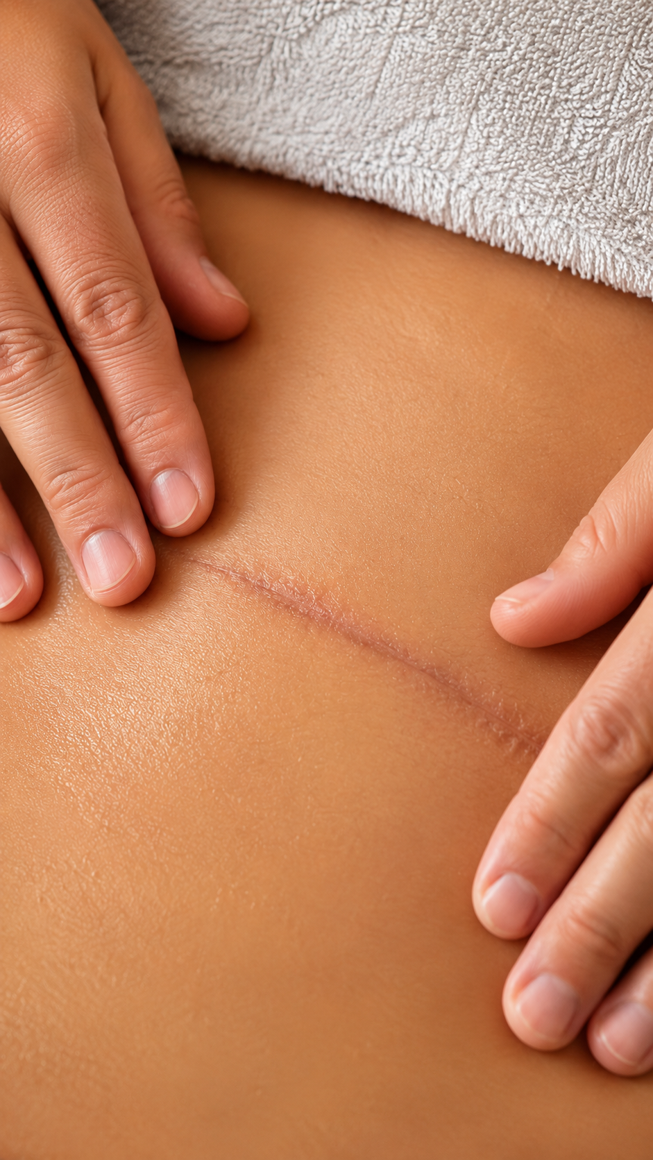
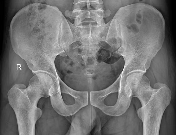
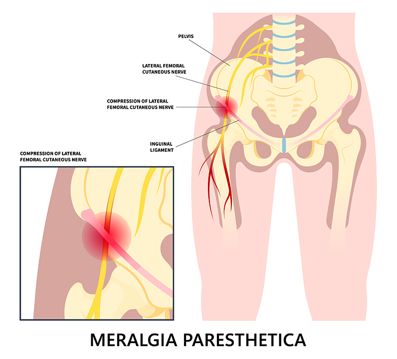
Meralgia Paresthetica is a condition characterized by numbness, tingling, and burning pain in the outer thigh. It occurs when the “lateral femoral cutaneous nerve” (LFCN), which supplies sensation to the skin of the thigh, becomes compressed or irritated. While not life-threatening, it can be uncomfortable and disruptive to daily activities. Now let’s see how Myotherapy treatment can help with this presentation. Causes of Meralgia Paresthetica Meralgia Paresthetica presents with symptoms of lateral thigh weakness and numbness, which are caused by compression or the lateral femoral cutaneous nerve. This compression can have typical causes, which include: Tight clothing – Wearing tight jeans, belts, or shapewear can compress the nerve at the hip height. Obesity or weight gain – Excess weight puts pressure on the nerve. Pregnancy – The growing uterus may contribute to nerve compression. Prolonged standing or walking – Excessive movement can irritate the nerve. Injury or trauma – Previous surgeries, injuries, or direct impact to the hip area can damage the nerve. Diabetes – Diabetic neuropathy may increase susceptibility to nerve-related conditions. Inguinal ligament – The LFCN passes right under the inguinal ligament, which may create compression on the nerve itself. All those factors can compress the branch of the femoral nerve, which innervates the lateral portion of the thigh. The origin of the nerve is at the lumbar level L2/L3. Indeed, when a portion of the skin has altered sensation, it is often a peripheral compression that causes the symptoms. Symptoms of Meralgia Paresthetica The symptoms for Meraglia Parestetica often involved one leg only, as it is quiet uncommon to get compression bilaterally. Those symptoms include: Burning, tingling, or numbness in the outer thigh. The skin of the lateral thigh can also become very sensitive and painful to the touch. Sharp or aching pain that worsens with prolonged standing or walking. If the compression is due to organs or the inguinal ligament, movement can aggravate the presentation due to the tightness of the structure during movement. Increased sensitivity to touch in the affected area. Muscle weakness is not a symptom, as this condition affects sensation, not motor function. How can myotherapy treatment help individualise this presentation? As a myotherapist, I specialise in muscular skeletal presentations, and we focus on soft tissues. Through a series of assessments, we can determine whether the compression is peripheral or root nerve compression. Let’s see how. Medical History In the first step, we examine the medical history and physical examination, identifying risk factors and symptoms. Along with the physical examination, we examine the Myotome and Dermatome. Examination On top of active range of motion and another orthopedic test to rule in and out other possible presentations, there are some specific tests which we want to focus on, Myotome and Dermatome. The Myotome are resisted movement, like in this case, hip flexion, knee flexion, knee extension, where it would result in positive findings if we have evident weakness and or back pain. This test would rule in a compression to the spine level. Dermatome, on the other hand, are used to test the connectivity of the cutaneous nerve. So with a sharp and soft object, we will mark some line along the thigh area, looking for loss or confused sensations. This test would rule in a peripheral compression of the nerve. Notice that both presentations can be presented at the same time. Other tests that can be done for this presentation include: Electromyography (EMG) – To rule out other neurological disorders. Imaging tests (MRI, X-ray, or ultrasound) – Identifying structural issues or nerve compression. For those tests, Giovanni would write a referral letter for your GP. Treatment Options for Meralgia Paresthetica As often happens, the treatment options are multiple and must be embraced in groups, not individually. The overall aim of any treatment is to relieve pressure on the nerve and reduce symptoms. Here is a list of treatment options and modalities: Lifestyle Modifications Wear loose-fitting clothing to reduce nerve compression. Weight management to decrease excess pressure on the nerve. Avoid prolonged standing or walking if symptoms worsen. Medical Treatments Pain relievers – NSAIDs (like ibuprofen) or acetaminophen for mild pain relief. Myotherapy treatment – along a series of myotherapy sessions we can reduce symptoms and improve the presentation. Corticosteroid injections – Reduce inflammation and pain. Nerve blocks – In severe cases, numbing the nerve can provide relief. Surgical Options (For Severe Cases) Nerve decompression surgery – Relieves pressure on the nerve. Neurectomy – Removing the affected nerve if pain is persistent. How Myotherapy Can Help At Melbourne Massage and Treatment, during a myotherapy session, after ensuring we are dealing with a Meralgia Paresthetica I may use a series of techniques to help you out with your symptoms. What technique to use is based on your individual presentation,, other cohexsitng presentations, adn also your choice and comfort. Here is a list of modalities used during a Myotherapy session: Muscle Energetic Technique (MET) – Helps reduce tension in the hip, thigh, and lower back muscles that may be contributing to nerve compression. Trigger Point Therapy – Addresses myofascial trigger points that can exacerbate pain and discomfort. Mobility and Strengthening Exercises – Improves mobility and reduces pressure on the nerve. Postural Education – Helps correct movement patterns that may be aggravating symptoms. Joint Mobilization – Enhances circulation and reduces inflammation in affected joints. Dry needling – Using a needle can help reduce pain and muscle ache and increase the neurological connection of those same soft tissues. After the hands-on treatment, we will then look into exercises that can help maintain the change we created. That said, there are other precautions to take in consideration, like: Maintain a healthy weight to prevent excess pressure on the nerve. Choose comfortable clothing that doesn’t constrict the waist or thighs. Incorporate gentle mobility and exercise into your routine. Monitor underlying conditions, such as diabetes, to reduce nerve-related complications. Conclusion Meralgia Paresthetica can be managed effectively with lifestyle changes, medical treatment, and preventive care. Myotherapy can be a valuable complementary […]