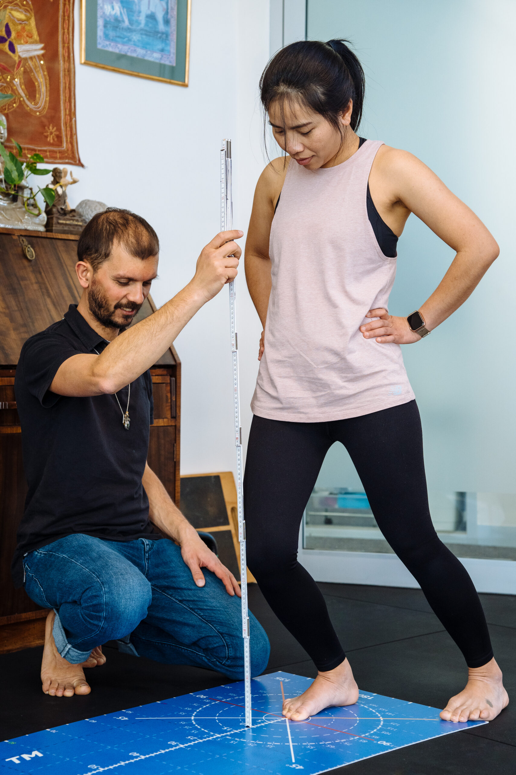
ACL stands for anterior cruciate ligament, and is the strongest ligament in the knee. ACL injuries can be time-consuming, depending on the severity. At Melbourne Massage and Treatment in Fitzroy North, I offer an ACL rehab program that aims to help you recover based on your clinical history, daily activities, including your sports, if any, and consider that the healing time can’t be boosted, but assisted. ACL – What It Is? ACL is the strongest ligament in your knee, and connects the femur to the tibia. Its role is to keep the knee stable, especially during activities that involve sudden stops, changes in direction, or jumping. You can then understand why so many athletes or sports lovers end up with an ACL-type injury. As with all ligaments in our body, the ACL doesn’t have an excellent blood supply, which makes its recovery longer, compared to what can be a muscle injury. What is Myotherapy and How Can It Help with ACL Rehab? Myotherapy is a hands-on treatment method that targets muscular pain, improves joint mobility, and addresses the root causes of musculoskeletal injuries. For ACL rehab, this approach is essential because an ACL injury affects not just the knee but the surrounding muscles, joints and movement patterns. Hands-on treatment can indeed be a good start for rehab and injury, as it not only creates a trust between the patient and the therapist, but can help in creating better awareness of the muscles, which in a second phase of the myotherapy appointment are going to be used for delivering tailored and injury-specific exercises. The goal is to support your ACL recovery and knee rehabilitation holistically. ACL Injury: Scan and Manual Test There are several methods of diagnosing an ACL injury. Scans: Arthroscopy – A keyhole surgery, where a camera is inserted in the knee to check the level of injury. Partial or complete ACL tear. Magnetic Resonance Imaging (MRI) – Less invasive than an Arthroscopy, but still expensive to run. Indeed, it is rare nowadays; in Australia, you can get an MRI prescribed for this presentation. More information is available here. Manual Test Lachman test Lateral Pivot Shift Test Anterior Draw Test Those together with an attemptive clinical history, range of motion and functional test would help to tailor a conclusion on a possible ACL injury. ACL and Surgery – Is Surgery Still a Thing? Surgery techniques and technologies have come a long way from what we could expect, and yes, ACL surgery is still a thing. That said, before going for surgery, you will be asked to try and recover from your injury with conservative treatment, like a rehab program. The reason behind this is: Suregey cost and pressure on the public health system Rehab gives as good results as surgery most of the time Surgery also goes by the severity of your injury – is any other knee tissue/structure injured, or is it only the ACL? Post surgery, you still have to go for rehab, and it would still take 6 to 12 months for a full recovery. Athletes may benefit the most from ACL surgery, given its longer-lasting effects on knee stability. More information about ACL rehab vs ACL surgery is available from the systematic review by Papaleontiou et al (2024). The ACL Rehab Phases: Strengthen Beyond the Knee To recover from an ACL injury, we need to look behind the mechanics of your lower limb, and set a starting and and ending point of recovery. Acute Phase In the acute phase, which is the first one to two weeks after injury, we may focus on isometric holds, gentle movement, and spend more time on hands-on treatment. In this phase, the aids of crutches is mandatory. Strength Phase Past the acute phase of the injury, you will notice how it would be easier to engage the knee joint, as the swelling would be eased, and therefore, you can achieve greater movement. There, we will start incorporating eccentric and concentric loads, with some light-weight or light-resistance bends. This phase can last somewhere between 10 and 12 weeks. All of these would be a step-by-step process. So the loads would be progressed weekly, based on the strength and mobility outcome. Sometimes, you will have to push through some mild pain, but the most important thing is that you keep that joint moving, and have good rest, good food too, to enhance the recovery at its best. Return to daily activity – RDA In the final stage, the focus would shift to plyometrics, an essential part of ligament rehab. That’s where we would re-train your knee to act pre-injury time. This can take another 12 to 24 weeks. It all depends on the severity of the injury and what your needs, in terms of sport or daily activities, are. So, the combination of myotherapy treatment with targeted rehabilitation exercises and fitness classes for recovery is what makes Melbourne Massage and Treatment service stand out. In my rehab-focused classes, we work on: Strengthening the muscles that support the ACL Improving balance and stability to prevent re-injury Gradual conditioning to safely return to sports, running, or everyday activities My Personal Approach At Melbourne Massage and Treatment, I work one-on-one with each client. I assess your movement, identify areas of weakness or tension, and design a personalised ACL rehab program. To be even more specific to your individual presentation, I do use THE MAT as an aid. A measurement tool that allows me to test your mobility and stability, giving us objective numbers and data to work on, too. Recovery from an ACL injury takes time, but with the right approach, you can regain full function and return to the activities you love. I’m here to guide you through every step of your ACL rehabilitation journey. If you’re ready to start your ACL rehabilitation in Fitzroy North or want to see how myotherapy can help, book a session with me at Melbourne Massage and Treatment today. Let’s get you moving stronger and safer than […]
Tag Archives: scan
Blog
When You Should Stop Running? And For How Long?
Here at Melbourne Massage and Treatment, Myotherapy Clinic in Fitzroy North, when treating patients who [...]
Continue reading
Blog
Pillow For Neck Pain: What You Need To Know
When treating someone for neck pain, a common question I get asked is: “Should I [...]
Continue readingJun

Blog
Femoroacetabular Impingement (FAI): What You Need to Know Before It Becomes a Bigger Problem
Hip pain can be frustrating, and not all hip pains are the same. I personally [...]
Continue readingJun

Blog
Femoral Anteversion: Why Your Hip Anatomy Changes the Way You Squat
“Oh, I can’t squat that deep, ” is what I sometimes get told by my [...]
Continue readingJun

Blog
Simple thing you can do for your Lymphatic System Well-Being. Go For A Walk!
As a therapist who offers Manual Lymphatic Drainage in Melbourne, I am blown away by [...]
Continue readingMay
Mar
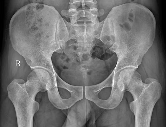
Musculoskeletal pain can be complex, and orthopedic tests and hands-on treatment, sometimes, can be a limited tool to individualise what is happening with the body’s internal structure. Indeed, there are times when a deeper look is required to ensure we are on the right path. This is where body scans imaging comes into play to identify presentations like tendinopathy, bursitis, ligament tear or other underlying conditions. The Role of Body’s Scan in Diagnosing Pathology Body scans include a series of imaging technologies, such as ultrasound, x-ray, MRI, to name a few. Ultrasound is a highly effective imaging tool used to assess soft tissue structures in real-time. Unlike X-rays, which primarily show bone, ultrasound provides detailed images of muscles, tendons, bursae, and ligaments. This makes it an excellent tool for diagnosing conditions such as: Tendinopathy – A chronic condition involving tendon degeneration due to overuse or injury. Bursitis – Inflammation of the bursae, the small fluid-filled sacs that reduce friction between tissues. Those tissue types are found along different body joints, like the shoulder and the hip. Ligament Tears – Partial or complete tears of ligaments, often occurring after trauma or excessive stress. Baker’s cyst – is a fluid-filled swelling that forms behind the knee, often resulting from knee joint conditions like arthritis or meniscal tears, causing discomfort and limited mobility. When we are suspicious of one of those presentations, due to positive results obtained by orthopedic test and medical history, including mechanism of injury, we attempt a recovery process, based on the type of injury, symptoms, and other relevant information. Along this recovery process, we may start with isometric exercises. If, with the first 6 weeks, and a series of sessions, 3 to 4 sessions with this time frame, we still don’t see a major recovery, then we may want to get extra investigation ongoing via an ultrasound scan, which can clarify the underlying pathology. It allows us to confirm or rule out certain conditions, ensuring that treatment strategies are aligned with the actual tissue damage (if any is present). On the other hand, based always on the individual case, we could also require X-rays, which are often more helpful in diagnosing conditions related to the bones, such as arthritis or fractures, as they provide a clear view of bone structure and joint spaces. MRI is a scan that is used for Brain imaging, and when the investigation needs higher details, like when looking at the spine or a joint that via ultrasound was not giving any sign of issue. Ultrasound is also comparable to MRI, as it is faster, easier to deliver, and has fewer complications. How can myotherapy treatment help recovery from what a body scans would show? As we already discussed in another blog, Myotherapy is a practice that looks into the well-being of the skeletal muscle structure. To understand what can be done about a painful presentation, we would initially take a detailed clinical history, then look into objective measurements, such as your movement and body presentation. Given the result we can obtain, we would build up a treatment plan which includes: Hands-on Treatment – Techniques such as deep tissue massage, myofascial release, and dry needling can help reduce pain and improve mobility. Exercise Prescription – Strengthening and mobility exercises help restore function and prevent future injuries. Load Management Strategies – Proper guidance on activity levels ensures tissues heal without excessive strain. That management technique would then be combined and adjusted around the scan’s results. Here are a few examples: Bursitis: If a bursitis is confirmed, medications may be given to reduce the inflammation of the bursa, for that, we concentrate on MLD treatment to further reduce the inflammation and exercises to build strength on the structure that needs support. Ligament tear: When talking of ligament tear, the healing time can dilagate to months if not also a year, so we know now why the 6 weeks program may was not as responsive. We will keep focusing on the strength of the muscle that surrounds the specific joint, and use hands-on treatment to boost blood to the area affected. Arthritis: Medication or dietary change may be put in consideration for pain management and inflammatory reduction. Also in this case, MLD can be used to manage the pain response, and exercises for mantain movement in the affected joint/s. When Should You Consider an Ultrasound or other body scans? If you experience ongoing pain, swelling, or restricted movement that is not improving with therapy, an ultrasound or other scan helps identify the cause. This can prevent prolonged discomfort and allow for a more targeted treatment approach. At Melbourne Massage and Treatment, in Fitzroy North, we aim to provide the most effective care possible. If you’re dealing with persistent musculoskeletal pain, book a consultation with Giovanni today. Together, we’ll determine the best action to get you back to optimal function. Frequently Asked Questions (FAQs) About Musculoskeletal Pain and Body Scans Imaging 1. What are body scans, and how do they help diagnose musculoskeletal pain?Body scans include imaging technologies such as ultrasound, X-ray, and MRI. These scans help diagnose soft tissue injuries (like tendinopathy, bursitis, and ligament tears) or bone-related conditions (such as fractures or arthritis). They provide a clearer picture of what might be causing pain, inflammation, or restricted movement. 2. Why is ultrasound commonly used in diagnosing soft tissue injuries?Ultrasound is highly effective for real-time imaging of soft tissues like muscles, tendons, bursae, and ligaments. It helps diagnose conditions such as tendinopathy, bursitis, and ligament tears, providing a dynamic view of the area being studied without the need for invasive procedures. 3. When should I consider getting an ultrasound or other scans for my injury?If you’re experiencing persistent pain, swelling, or limited mobility that isn’t improving with initial therapy (such as exercises or hands-on treatment), it might be time to consider an ultrasound or other scans. These can help identify the underlying cause of your symptoms and allow for a more targeted treatment approach. 4. How do orthopedic […]