
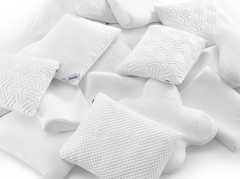
When treating someone for neck pain, a common question I get asked is: “Should I change my pillow?” or “I slept badly, my pillow is not good”. In fact, there is a common belief that a pillow or a bad night’s sleep is the cause of constant neck pain. While pillow comfort matters, clinical experience in myotherapy in Melbourne shows that pillows are rarely the root cause of ongoing neck pain. At a clinical level, neck pain is usually driven by: movement dysfunction poor spinal cervical stability previous injury history and reduced muscular control Therefore, a good pillow can support symptoms, but it does not fix the underlying issues that drive your pain. Coburg Myotherapy Insight: What Actually Causes Neck Pain? At Melbourne Massage and Treatment, a myotherapy clinic in Coburg, I consistently see neck pain linked to five key factors. 1. Spine Mobility and Stability Dysfunction The neck’s vertebrae are part of a full kinetic chain involving the thoracic spine, shoulders, and rib cage. Under this aspect, indeed, we should look into the lack of thoracic mobility which may cause the stable portion of your cervical (C3-C7) to seek that mobility capacity. But as we well know, a stable joint can’t act as a mobile, and vice versa. In more detail, poor spinal mechanics lead to: muscle overload in the neck joint irritation tension during rest and sleep And about the pillow, it is an object that cannot restore spinal movement or control. Isn’t it? 2. Whiplash History and Incomplete Rehabilitation Other patients with chronic neck pain may have a history of whiplash that was never fully rehabilitated. Indeed, a whiplash accident, as a result of a car crash or even during a contact sport incident, can lead to chronic neck pain, especially if not rehabilitated correctly. This happens because during a whiplash, the cervical joints get put under extreme force and can lead to a torn ligament or laxation. However, without structured rehab, long-term changes in: deep neck flexor control proprioception postural endurance can persist for years, or even show up after years of post-injury. So again, no pillow can change these symptoms or help you recover from such an injury. 3. TMJ Dysfunction and Jaw Tension Temporomandibular joint (TMJ) dysfunction is another underlying cause of neck pain. By following the kinetic joint chain we discussed earlier, we see that the TMJ is the next joint in the chain, after the cervical one. Therefore, Jaw clenching and TMJ irritation can: increase suboccipital muscle tension contribute to forward head posture amplify cervical joint stress That’s where you may wake up with more neck pain than during the daytime. The clantching can be heavily responsible for that. This is why neck pain is often worse under stress or during sleep. 4. Hypermobility and Joint Instability Following the concept of a torn ligament, as per a whiplash incidence, joint hypermobility is clearly another underlying issue for neck pain. This happens because there is a lack of stability and control in those cervical ligaments. This leads to: reliance on passive ligament support early muscle fatigue poor tolerance of sustained sleeping positions A “supportive pillow” alone may not solve instability and can sometimes reinforce dependency on external support rather than active control. 5. Neck Strength and Motor Control (8–12 Week Rehab Window) In my experience, this is the big issue. Most people don’t even think about the strength of their neck or upper shoulder muscles, and this is where things can go really wrong. Indeed, one of the most effective long-term treatments for neck pain is a structured strengthening program that targets: deep cervical flexors scapular stabilisers postural endurance muscles As per many exercise programs that intend to strengthen the body structure, we look at 8–12 weeks of progressive exercise-based rehabilitation. This is why many patients searching for neck pain treatment in Melbourne benefit more from rehab than passive support strategies. What the Research Says About Pillows and Neck Pain To support with evidence, why a pillow is not going to change in the long term, your neck pain, we can look at a systematic review from Pang J. et al. (2021), which found: Some pillow designs may reduce symptoms But long-term structural or functional improvements are limited and inconsistent Therefore, pillows may improve comfort, but they do not fix the underlying cause of neck pain. And yes, the pillow consistency can make a difference, but, again, it does not fix the neck pain, nor would it cause neck pain. Melbourne Myotherapy Approach: What Actually Works At Melbourne Massage and Treatment – Myotherapy Clinic in Coburg, I focus on evidence-based management of neck pain. Indeed, my approach to a client who presents with neck pain is structured as follows: Detailed analysis of clinical history – including previous history of injury and past/current sport activities Clinical assessment – active and passive range of motion, and neurological test where/when needed Outline your short-term and long-term treatment – what you want to achieve in today’s session and in the upcoming weeks/months, and what you are ready to do about it (exercise-wise) Treatment plan outline – what I believe could be a tailored treatment plan given the results of the above findings, including your short/long-term goals In all of these, Remedial Massage plays a crucial initial role to relax the nervous system, reduce initial pain and discomfort, promote healing and movement, but even techniques like mobilisation are ideal for treating neck pain and improving the initial outcome. But again, hands-on treatment is only the first step of the journey, and along with other myotherapy sessions that follow the initial consultation, exercises have to be the main focus. Exercises for the neck, for the shoulders and for your back. In summary, here is a dot point list of what a treatment plan aims for: cervical + thoracic mobility restoration deep neck flexor strengthening scapular control TMJ and jaw tension management (if needed) graded load tolerance programs These interventions address the true drivers of pain rather than just […]
Monthly Archives: June 2026
Blog
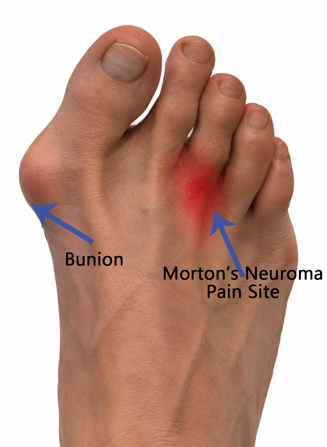
Bunion and Morton’s Neuroma: Can a Bunion Affect Neuroma Recovery?
While a bunion and a Morton’s neuroma are two different foot conditions, if they are [...]
Continue readingSep
Blog
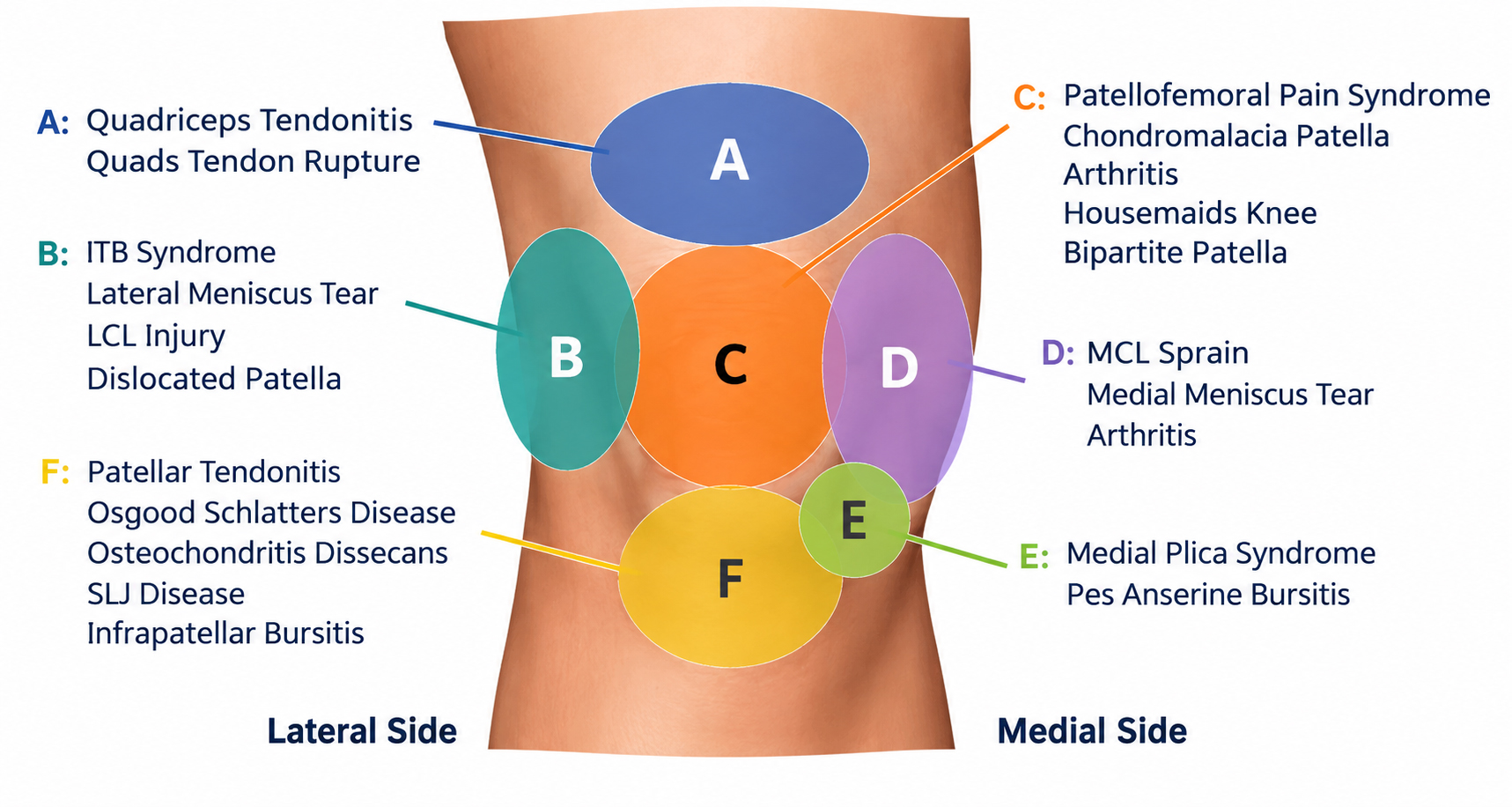
Knee Pain in Coburg
Knee pain is a common form of injury and can affect people of all ages [...]
Continue readingSep

Blog
Post-Surgery MLD Melbourne – Vodder MLD Therapist
Any form of surgery is an invasive procedure for the body to deal with, and [...]
Continue readingSep
Blog
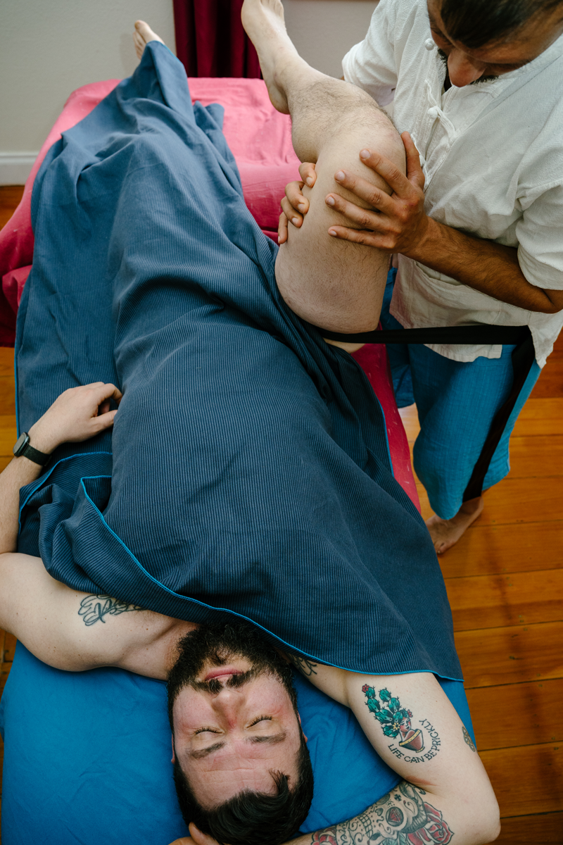
Hip Pain in Coburg: Causes, Symptoms & Treatment
In the many presentations I get to see here at Melbourne Massage and Treatment in [...]
Continue readingSep

Blog
Lymphatic Walking: Let Me Tell You Why It Is Another Trend
Lymphatic Walking is another trend that you really don’t need to overthink. But let me [...]
Continue readingAug

Jun
Hip pain can be frustrating, and not all hip pains are the same. I personally experienced Femoroacetabular Impingement (FAI) pain myself, and it is not fun. Indeed, this type of pain can stop you from training, affect your sleep, make sitting uncomfortable, and eventually impact your quality of life. Over the years, I’ve seen many active people spend months treating the symptoms without understanding the actual cause. If you’re experiencing groin pain, hip stiffness, pinching during squats, or discomfort after prolonged sitting, FAI may be worth investigating. What Is Femoroacetabular Impingement? In simple terms, Femoroacetabular Impingement (FAI) occurs when there is abnormal contact between the femoral head (the ball) and the acetabulum (the socket) of the hip joint. As with any tissue that gets overstimulated, this can result in irritation and inflammation, leading to damage to the labrum, cartilage, and surrounding tissues over time. In some cases, if left unmanaged, it may contribute to the development of early hip osteoarthritis. The symptoms that most people come up with when experiencing FAI are: Deep groin pain Hip stiffness Clicking Catching Locking sensations Pain when sitting for extended periods. Those symptoms are typically aggravated by activities involving Deep hip flexion Squatting Running Kicking Cycling Getting in and out of a car. The Different Types of FAI As we have already seen in the antirotated and retroverted hip presentations, we are all different, and even a Femoroacetabular Impingement can present differently. Let’s have a look at the different types of Femoroacetabular Impingement. Cam Impingement Cam impingement occurs when the femoral head is not perfectly round. During hip movement, particularly flexion and internal rotation, the abnormal shape creates increased pressure against the edge of the socket. This is the most common form seen in young athletic populations. Pincer Impingement Pincer impingement occurs when the acetabulum provides excessive coverage over the femoral head. The socket effectively “overhangs,” increasing the likelihood of compression during movement. Mixed Impingement Mixed FAI is the most common presentation clinically. In this situation, both cam and pincer characteristics are present simultaneously, resulting in a combination of abnormal contact from both the femur and the acetabulum. Who Is More Likely to Develop FAI? The common ground for an FAI presentation includes young and middle-aged active individuals. A higher prevalence is seen among athletes participating in sports that involve: Repetitive hip loading during adolescence Football Hockey Soccer Martial arts Dance Running. This evidence shows that sports activities play a crucial role in FAI presentation, but it is also important to consider that genetic factors could be involved. On the other hand, as per any physical structural presentation, not all the people who present with a FAI may experience pain. Many people have structural changes visible on imaging but remain completely symptom-free. Is a Scan Worth It? This is one of the most common questions I get asked when someone presents with some sort of ongoing pain or even acute discomfort. My answer is usually: it depends on your symptoms and how long they have been lingering around. Why I don’t recommend a scan as a first thing to go for (unless I am suspicious of something that I can’t treat directly) is because a scan may find an abnormality in the body, but that doesn’t mean that what we see is actually the source of the pain. In fact, many people may have a FAI presentation and have no symptoms at all. Therefore, a scan alone should never determine treatment decisions. Sp, a diagnosis of FAI should combine: Clinical history Physical examination Symptom presentation Imaging findings So yes, a scan should support the diagnosis, not create it, or it would be really chaotic to define why someone is experiencing pain, and create a treatment plan for it. If You Need Imaging, Which Scan Is Best? Step 1: X-Ray For most people, a standard pelvic and hip X-ray is the first and most appropriate imaging investigation. X-rays are excellent for identifying the bony shapes associated with cam and pincer impingement and are considered the primary imaging modality in the assessment of FAI. The downside of X-rays is that they involve radiation, so if possible, avoid them. Step 2: MRI If symptoms are there for a prolonged period of time, and exercise therapy is failing to restore functionality, an MRI becomes extremely valuable. MRI can assess: Labral tears Cartilage damage Joint degeneration Other soft tissue causes of hip pain Many hip specialists consider MRI the cornerstone investigation when assessing intra-articular damage associated with FAI, given the high definition of the image and results. Step 3: CT Scan CT scanning is generally reserved for surgical planning or when a very detailed understanding of the hip’s bony anatomy is required. CT provides excellent visualisation of bone structure, but it is not usually necessary as an initial investigation. So, When Should You Get a Scan? In my clinical opinion, based on current evidence, imaging becomes worthwhile when: Hip or groin pain has persisted for more than 6–12 weeks Symptoms continue despite appropriate rehabilitation The range of motion is progressively decreasing Mechanical symptoms such as catching, locking or giving way are present, and are painful Surgery is being considered The diagnosis remains unclear after clinical assessment If your symptoms are mild and improving with treatment and exercise, I would not bother to get an image taken. Exercises are a great way to maintain hip pain-free and keep your body going. Exercise Protocol for FAI This is where we need to put some focus: Exercises! So, first things first, when someone presents with a FAI, we want to take away or modify the habit that we can, in order to reduce discomfort and hip pain. An example could be removing squat from an exercise program or reducing the time spent in a seated position. Next, we would start looking into your mobility capacity at the ankle level, especially if you are someone who runs as part of a sports activity. And all of this is part of a protocol […]

Jun
“Oh, I can’t squat that deep, ” is what I sometimes get told by my clients when I train them for exercises. What do I think about it? I think they are right, maybe they can’t, and that’s ok. And why can’t they? Well, we are all different, and not one squat is equal to the other one. And possibly the answer is behind the fact that they may have a femoral anteversion. Indeed, femoral anteversion can affect how your hips rotate, how your feet naturally position themselves during a squat, and even how comfortable certain exercises feel. We Are Not Built the Same. And That’s Fine! Most recently, I was working with a client who presented with Lipoedema and, consequently, hypermobility, and when we got to work on her squat we notice that deep squat for her was not a thing (even thought she is hypermobile). Her PT, on the other hand, was asking her to just keep trying, gave her an app-video to train with, and told her that the squat had one way to be. Obviously this is not the case. We all squat differently, and there is nothing wrong with it. Then, after a short investigation, we realise that she can go deeper in the squat if she uses a few tricks and tips. This is because her hips are antroverted. Moving forward from this single case scenario, we also have to remember that our movement is influenced by: Muscle strength Mobility Motor control Previous injuries – (actually, she also had severely injured her L ankle when she was a teen, and her dorsiflexion is compromised on that side) Joint structure Bone morphology The last point is often overlooked. In fact, the shape and orientation of the femur can significantly influence how the hip moves. This is where femoral anteversion and its counterpart, femoral retroversion, become important. Therefore, understanding these anatomical differences can help explain why you squat comfortably with feet narrow, standing and facing forward, while another naturally prefers a wider stance with their toes turned outward. What Is Femoral Anteversion? Femoral anteversion refers to the forward orientation of the femoral neck relative to the shaft of the femur. More simply, this means that the head and neck of the thigh bone are rotated more anteriorly than the norm. In fact, everyone is born with some degree of femoral anteversion. What can happen is that during growth and development, the amount gradually decreases, but the final angle varies considerably between individuals. Research by Scorletti M et al. (2020) has shown that femoral versions are present on a spectrum, where there is significant variation even among healthy adults. That’s where the word “normal” has no application. There is no “normal hip”. What indeed is normal is the variability of human anatomy. Regarding the characteristics of people with greater femoral anteversion, typically, we find: Increased hip internal rotation Reduced hip external rotation A tendency toward a more forward-facing foot position Greater comfort in certain squat positions Different movement strategies compared to those with retroverted hips And therefore, let’s underline the fact that femoral anteversion is not a pathology, but it is simply an anatomical variation. What Is Femoral Retroversion? Now that we have looked at femoral anteversion, it is time to look at the opposite presentation. In this case, the Femoral retroversion occurs when the femoral neck is oriented more posteriorly relative to the femoral shaft. These individuals often display: Increased hip external rotation Reduced hip internal rotation A natural toe-out posture Preference for wider squat stances Reduced comfort with feet pointing straight ahead None of those presentations is better than the other one, or more “normal”. They are simply different anatomical variations of the hip biomechanics. The result of one or the other is that, based on the presentation you show up with, there are going to be certain movements that are easier or less easy for you. The real issue stands when people attempt to force a movement pattern that doesn’t match their anatomy, especially if they told: “this is how you squat/move”. How Femoral Anteversion Influences Your Squat Now that we have a better idea of what is what, in terms of hip anatomy variation, we can look at how femoral anteversion affects your squatting. As the hips flex during a squat, the femoral neck moves within the acetabulum (hip socket). The available space for movement depends partly on the shape and orientation of the bones involved. Remember, the femoral hip joint is a socket/ball joint, so a sphere shape (femoral head) rolling inside a concave socket. So, if you are one of the individuals who present with greater femoral anteversion, you will find it more comfortable doing movements where: Feet are relatively straight Your stance is narrow to moderate Knees track naturally over the feet Don’t need an excessive toe-out stance Indeed, by contrast, individuals with femoral retroversion often prefer: A wider stance Greater foot turnout More externally rotated hip positions And again, this is why two healthy people can perform completely different-looking squats and both be moving optimally for their anatomy. Trying to force everybody into the same squat position ignores the reality of individual biomechanics and would put one or another individual in a place of lack of confidence or body negativity. Why Foot Position Matters So, now that we see both anteversion and retroversion hip presentation, it’s time to understand why the feet position can make a difference when doing a squat, and where it is needed for a deeper squat and where it is optional. Indeed, as mentioned above, for someone with a retroverted hip, having feet straight and worst, even if too close to each other, and delivering a squat, is not a thing. This is dictated by the fact that the foot direction, such as straight or laterally directed, is given by the rotation that we apply at the hip joint. A person with significant femoral anteversion may naturally feel strongest with minimal toe-out, whereas someone with retroversion […]