It seems we can’t find what you’re looking for. Perhaps searching can help.
Category Archives: Melbourne Massage and Treatment
Melbourne Massage and Treatment is a home-based massage studio run by Giovanni, a qualified Myotherapist passionate about health and healing.
Melbourne Massage and Treatment Services.
Giovanni started in Massage career back in 2016 at the SSNT in Fitzroy.
After that, he worked and travelled in Asia and Europe, specialised in Thai Massage and Applied Manual Lymphatic Drainage (MLD), and completed a diploma in Remedial Massage and Advance Diploma in Myotherapy.
Giovanni is finishing a Bachelor in Health Science, Myotherapy Clinic at Torrens University.
What type of condition does Giovanni treat?
Plantar Fasciitis
Frozen Shoulder
Lower Back Pain
Neck Pain
Head Aches
Fibromyalgia
Knee Pain
Carpal Tunnel
Tennis Elbow
Golfer Elbow
Achilles Thenopathy
Muscle tear
Recovery from bond fracture
Recovery from Surgery (MLD)
TMJ pain
Preeclampsia
Improving mobility
What is this blog About?
Melbourne Massage and Treatment blog is about Giovanni’s experience within the massage industry, health topics, and an explanation of how different massage services works.
Giovanni likes to ensure his client knows what happens to their body when they are in pain or living with discomfort. He believes body awareness is vital to the recovery/healing process.
If you are looking for treatment, book your next appointment with Giovanni now.
Or if you have any questions, get in touch now.
Massage Near Me – Coburg
Melbourne Massage and Treatment studio is located on Blair St, Coburg, 3058.
Easy to get here by public transport, like tram 1, 6 or 19, or by train, as Giovanni lives nearby Moreland Station.
Also, if you need to drive here, plenty of car park spots are available along Blair St.

Blog
Why the Gluteus Medius Is Essential for Balance, Longevity and Fall Prevention
One thing that can really create terror while we age is falling. No matter the [...]
Continue reading 16
Mar
Mar
Blog
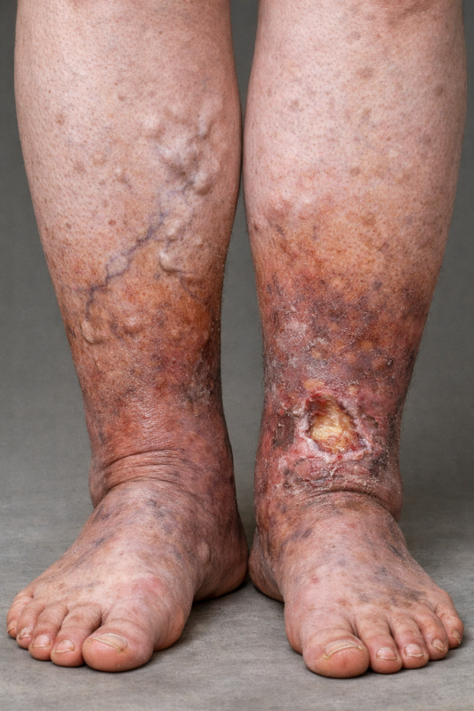
Chronic Venous Insufficiency and Swollen Legs: Compression Is The Way To Go
In Australia, Chronic Venous Insufficiency (CVI) affect more women than men, with a ratio of [...]
Continue reading 09
Mar
Mar
Blog
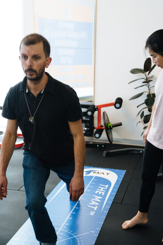
It Is Never Too Late To Join A 1:1 Fitness Class
At Melbourne Massage and Treatment in Fitzroy North, as a clinical Myotherapist, I am enthusiastic [...]
Continue reading 02
Mar
Mar

Blog
MLD vs Lymphatic Massage: A Clinician’s Guide to What Works and Why
If you’re searching for lymphatic therapy in Fitzroy North, it’s common to see terms like [...]
Continue reading 23
Feb
Feb

Blog
Shoulder Pain Isn’t Just a Shoulder Problem
Shoulder pain is one of those presentations that can stop you from enjoying your day. [...]
Continue reading 16
Feb
Feb