
Experiencing a vertebral fracture can be an overwhelming and challenging experience to recover from, but this doesn’t mean there is no safe protocol and successful treatment pathway out there. At Melbourne Massage and Treatment, I am here to assist you in this complex journey, which could be by offering MLD treatment, Myotherapy or Fitness Class. But let’s first understand what fractured vertebrae mean, and what we have to be aware of when working with this type of injury. Spinal Damage vs. No Spinal Damage Let’s start to look into what difference makes to have a spinal fracture where the spinal cord was injured and where it was not. With spinal cord damage, a fracture may injure the spinal cord or nerves, leading to severe symptoms such as numbness, weakness, or paralysis. These cases are medical emergencies requiring hospital care. The rehabilitation process for someone who encounters spinal damage varies based on the severity of the injury. Surgery may be necessary to repair the nerve, but there is also the fact to consider that there may not be a recovery option and life paralysis (quadriplegic or paraplegic) as an outcome. Without spinal cord damage, it is a result of a bone fracture only, without affecting the cord. These are painful but often managed with an initial period of rest and bracing and gradual rehabilitation. At our Fitzroy North clinic, Giovanni carefully assesses your needs and works alongside your medical team to provide safe and effective rehabilitation. Cervical, Thoracic, and Lumbar Vertebrae Your spine has three main regions, and fractures behave differently depending on location: Cervical (neck): Mobile but delicate; fractures here can have severe consequences. Thoracic (mid-back): Stabilised by the rib cage, but injuries here often come from higher-energy impacts. Lumbar (lower back): These vertebrae carry the body’s weight, so fractures here cause significant pain and restricted movement. Based on where the fracture is, the treatment and recovery options and plans differ. Scans for Diagnosis To properly understand the type of fracture and the severity of the fracture itself, scans are essential. Here is a short list of what diagnostic scans are available and which are most commonly used, and why: X-ray: The first step to confirm a fracture. This type of test is good to see the fracture at the bond level; it is quick, but as a downside, it exposes you to radiation. CT scan: Provides detailed 3D imaging to assess the fracture’s stability. The downside of a CT scan is that, as it is based on X-Ray technology, it can still expose you to radiation, and it can take longer to be delivered, and it is essential to be lying down while receiving the scan. MRI scan: Compared to X-Ray technology, MRI scan would not expose you to radiation, and is used to detect any involvement of nerves, discs, or the spinal cord along the fracture, as this type of scan is used for water-based tissue in the body, and not bones. These scans help guide safe rehabilitation, ensuring the right treatment approach from day one. Something else to keep in mind from the result of the scan is that not everything that a scan shows must impact your life. Indeed, a building disk may show in your scan, but that doesn’t mean that that specific pathology is something related to your spine fracture (it may have been there already before), and that doesn’t mean the body would not look after it while you are recovering from the spine injury. Types of Vertebral Fracture Common fracture types include: Compression fracture – vertebra collapses, often linked to osteoporosis (also called a wedging fracture). Burst fracture – bone shatters outward, sometimes threatening the spinal cord. Flexion-distraction fracture – usually from high-speed accidents where the spine bends suddenly. Fracture-dislocation – bone and soft tissues are displaced, often requiring surgery. Avulsion – It is a type of stress fracture, characterised by a small piece of bone pulled away from the main bone by a muscle or ligament (typical along the transverse process). Mechanism of Injury Fractures can occur from: High-energy trauma – car accidents, falls, sports collisions. Low-energy stress – in osteoporosis, even coughing or bending can trigger a fracture. Scheuermann’s disease – in this specific condition, the vertebrae may grow at different heights compared to the sagittal plane. A meticulous clinical history intake can help in figuring out he chance of you suffering from a vertebral fracture. Healing Time and Recovery As per all non-complex bone fractures, most vertebral fractures take 8–12 weeks to heal, even if recovery varies depending on age, bone health, and whether surgery was required. What we know is that nothing can actually boost the healing, but different therapies, active and passive, can help in assisting the healing process, ensuring a positive outcome. What then can be done during the recovery time is: Early phase: Pain management and protection of the fracture. Rehabilitation phase: Gentle guided movement, strengthening, and improving mobility. With myotherapy support, clients can return to safe daily activities while minimising the risk of re-injury. What to Avoid in the Early Stages of a Vertebral Fracture As mentioned earlier, in the early stage of vertebral fracture, it is important to prevent further damage to the spine and wear a corset that helps in stabilising the spine, while the body is starting the calcification of the bone. Even though you may wear a support, you will want to avoid: Heavy lifting, twisting, or bending movements. Prolonged sitting without support. High-impact exercise or activities. Movement is still recommended, as it can still promote fluid movement and relaxation. Therefore, it is possible to go for walks, move your arms, and move your legs even if in a seated position. Manual Lymphatic Drainage Massage in the Early Phase of a Vertebral Fracture At Melbourne Massage and Treatment, I got to offer MLD as a form of treatment for relaxation, which can have a positive impact on pain perception and tension relief from the spine area. MLD is a gentle […]
Tag Archives: MLD

Blog
Vertebral Fracture in Fitzroy North: What You Need to Know
Experiencing a vertebral fracture can be an overwhelming and challenging experience to recover from, but [...]
Continue readingOct

Blog
Fibrosis After Cosmetic Surgery | Post-Surgery MLD Melbourne
Within the last few years, cosmetic surgeries have been on the rise in Australia. These [...]
Continue readingSep

Blog
How to differentiate Muscle Pain and Joint Pain – A Case Study
A pain response is a signal created by the brain to let you know that [...]
Continue readingSep

Blog
How Remedial Massage Reduces Stress?
Modern life places ongoing pressure on both body and mind, leaving many Australians searching for [...]
Continue readingAug

Blog
Thai Yoga: Mobility & Relaxation
Thai yoga combines assisted stretching with mindful breathing to restore mobility, release tension, and create [...]
Continue readingAug
Sep
Within the last few years, cosmetic surgeries have been on the rise in Australia. These types of interventions can be helpful for quick body changes. Still, the recovery process post-surgery is often under-estimated and misinterpreted, especially when, past a couple of days or just a few weeks, the body’s response to surgery leaves behind hard lumps, thick skin, and reduced sensitivity. The thought skin and lumps are simply fibrosis, and the reduced sensitivity results from damaged nervous system endings. At Melbourne Massage and Treatment in Fitzroy North, I specialise in Manual Lymphatic Drainage (MLD) using the Vodder technique, helping clients reduce swelling, assisting the recovery, and breaking down fibrosis post cosmetic surgery, safely and effectively. If you’re looking for post-surgery care in Fitzroy North or Melbourne, here’s how MLD can support your recovery and improve your results. What is Fibrosis After Cosmetic Surgery? Fibrosis formation post cosmetic surgery is the result of tissue damage that occurred during the surgery. When going for an intervention like liposuction, where fat is removed from the body (either at the abdominal level, arms or legs or elsewhere), the body, to replace the void left by the fat removal, builds up fibrotic tissue. The fibrotic tissue is mainly made of collagen. While this reaction is natural, it can often cause: Hard lumps or nodules under the skin; Uneven skin contour or texture; Tightness or restricted mobility; Tenderness or discomfort. There is no real way around those types of side effects post-liposuction, at least in the short term, and the body would take weeks to recover fully (up to 3 months). That said, everybody reacts differently to this type of intervention, and based on the type of intervention received, the recovery process can vary. Who Benefits Most From Lymphatic Drainage Massage After Cosmetic Surgery? Here is a short list of cosmetic surgery interventions that are going to leave you with fibrosis in the post-surgery time, and that would benefit from Lymphatic Drainage Massage intervention: Liposuction (abdomen, thighs, arms, chin) – This includes liposuction for Lipoedema management too. Tummy tuck – Either due to post-liposuction or from severe weight loss. Breast surgery (augmentation, reduction, reconstruction) – If this is due to breast cancer, one should be aware of any risk of Lymphoedema development. Facelifts and neck lifts Brazilian Butt Lift (BBL) – This type of intervention requires fat to be removed from other body parts, as the abdomen, and that’s where fibrosis would build up. How MLD Helps Reduce Fibrosis Manual Lymphatic Drainage (MLD) is a gentle, specialised technique that stimulates the lymphatic system to clear excess fluid, reduce swelling, and assist with the healing process. After surgery, your lymphatic system could be damaged and can struggle to keep up with its work, and that’s where MLD makes a big difference. Indeed, the stimulation of the Lymphatic System, via MLD therapy, can help in assisting your recovery and ensure that the fibrotic tissue gets absorbed and dismissed, restoring freedom of movement and leaving you soon after treatment in a deep relaxation state. I trained in the MLD with the Vodder style, therefore I can provide precise, tailored treatments that are safe for sensitive post-operative tissue. Book your post-surgery lymphatic drainage in Fitzroy North today to safely reduce fibrosis. When Can I Start MLD Treatment Post Cosmetic Surgery? Generally, MLD is safe to start as soon as the antibiotic cycle is ended post-cosmetic surgery. Given the light touch of this type of therapy, we aim to produce no pain during the treatment, so we can work close to the surgical side, without affecting the recovery process. On the other hand, I found myself occasionally referring patients to the local nurse or GP here at Fitzroy North Doctors, as their recovery immediately post-surgery was compromised by misleading suggestions and procedures offered by overseas cosmetic clinic surgery. If you are not sure about what’s going on with your recovery, please, before placing a booking for an MLD treatment, talk to your GP about your recovery state, and if you have any questions regarding MLD treatment, you can always reach out to me via the contact page. How Many MLD Sessions Do I Need To Reduce The Fibrosis? As mentioned earlier, everyone responds differently to cosmetic surgery, but in my experience, it would take at least 4 to 6 weeks to start seeing a significant difference in fibrous tissue presence. That said, the number of sessions and the time length of the sessions can vary, based on the area where you received the surgery. Abdominal surgery only: I will recommend 2 to 3 treatment per week, for the first 3 weeks. Within the first week, we may spend 1 hour per session, and from the 2nd week onwards, we reduce the treatment to 45 minutes. Multiple liposuction sites: as there are multiple areas where you received a surgery, we may need to extend the time of treatment up to 1 and a half hours initially, or go for multiple sessions, each for a different area. The first few treatment may take longer as we want to spend some extra time trying to break down the fibrosis with a gentler touch, due to the high sensitivity of the body, which is high due to post-surgery. As the sensitivity decreases, and we can apply further pressure, we can achieve the same result in fibrosis reduction with less time. If you are not sure what works best for you, you can book a 15-minute free online consultation, so we can discuss your needs and work out a treatment plan in accordance with them. MLD Prices in Fitzroy North All my services are offered at the same rate and are as follows: 90 mins – $175 1 hour – $125 45 mins – $ 115 30 mins – $90 All those prices are inclusive of GST. The 90-minute option is available only if required, and not via the booking system. Why Choose MLD at Melbourne Massage and Treatment in Fitzroy North? Not all lymphatic […]

Aug
As a Lymphoedema therapist, I often get asked what the difference is between Lymphoedema and Lipedema. In this blog, we will explore the differences, the similarities, and what can be done for prevention, management and treatment of those presentations. Furthermore, we will look into how Lipoedema can degenerate into a Lipo-Lymphoedema, and why this is not the case for everyone. What is Lipoedema? Lipoedema is a chronic adipose tissue disorder that primarily affects women. On a global scale, we know that about 11% of women are affected by this presentation, and it often runs in families as it has a strong genetic component. The major characteristics of Lipoedema are an abnormal and symmetrical accumulation of fat around the hips, buttocks, thighs, and legs, and upper arms. On the leg area, the fat appears in abundance in the medial side of the knee, too. Where feet are completely untouched by the fat accumulation, this fat is resistant to diet and exercise and is often painful to touch. The pain is due to the cutaneous nerve entrapped in the fatty tissue, and so delivers a pain response when stimulated. Other Lipoedema key features: Often triggered or worsened by hormonal changes Symmetrical fat distribution Soft, nodular, or lumpy tissue Pain and easy bruising – as per the pain, bruising is due to blood capillary compression from the fat, and so, is easily damaged by touch No skin thickening or pitting in the early stages Nowadays, there is increasing awareness about this presentation, and more and more women find benefit from a management protocol that is not only about cardio and exercise. Part of the Lipoedema management includes: Movement Compression stocking Antiinflammatory diet Skin care Where and if needed, cosmetic surgery intervantion What is Lymphoedema? Lymphoedema, on the other hand, is a condition where lymphatic fluid builds up in the tissues due to a malfunctioning lymphatic system, causing chronic swelling. Compared to Lipoedema, Lymphoedema is strictly related to the Lymphatic system. It can be primary (congenital or hereditary) or secondary (due to trauma, surgery, radiation, or infection affecting the lymphatic system). Lymphoedema characteristics: Unilateral or asymmetrical swelling (though it can be bilateral) Pitting edema – It consists of deep indentation (pitting) left behind on the skin when pressure is applied Skin changes over time (fibrosis, hyperkeratosis, papillomatosis) Affects feet and hands as well – primary lymphedema would start from the extremity Heaviness or tightness in the affected area – can potentially be pain-free, but the limb/s may feel very heavy It does affect men and women – only primary lymphedema has a genetic component Lymphoedema Management The management of Lymphoedema is more tricky than lipoedema, as everyone may react differently to the management, it can be related to other health issue which needs to be considered, and requires the patient to be active in the management side of things. At Melbourne Massage and Treatment, I treat different types of lymphedema, as per the upper and lower body, focusing on an initial reduction of the swelling via a combination of Manual Lymphatic Drainage (MLD) and compression with Combined Decongestive Therapy (CDT). The management of this presentation can take anywhere between 3 and 5 or more appointments, depending on the severity of the presentation. The treatments are better done in close proximity, 24 to 48 hours one after the other, so that we give no time to the body to accumulate fluid back under the skin. Once the combination of treatment allows us to achieve the desired result, which is bringing the limb/s to a thinner size, you will be scheduled for a custom garment wear compression, which will guarantee to maintain the results achieved. This is usually done at other clinics, like Sigvaris or Juzo clinics. Those clinics are specialised in the making of garment wear. Custom garments wear last about 6 months, so twice a year, you will need to change them, and if needed, because the limb/s may start swelling again (especially in summer, when there is a change of atmospheric pressure, due to the heat), a short series of MLD and CDT therapy may be needed. Key Differences between Lymphoedema and Lipoedema Feature Lipedema Lymphoedema Cause Abnormal fat metabolism Lymphatic dysfunction Gender prevalence Almost exclusively women Affects both sexes Onset Often at puberty, pregnancy, or menopause Can be congenital or triggered by injury/surgery Distribution Symmetrical, lower limbs and arms Can be asymmetrical; any body part Feet/Hands Spared Usually involved Pain Tender, painful fat Often painless, heavy feeling Skin texture Soft, nodular fat Skin thickens over time (fibrosis) Pitting Rare (early) Common (early) Response to elevation Minimal improvement Often improves with elevation (if early stage) Bruising Common Not typical Common Characteristics of Lymphoedema and Lipoedema As seen above, the characteristics of Lipoedema and Lymphoedema are different, but, both conditions share chronic swelling, potential functional limitations, and a need for long-term management: Both can cause leg discomfort, heaviness, and swelling Both may lead to reduced mobility Neither condition improves with calorie restriction or exercise alone – it is more about stop the intake of inflammatory food Compression therapy is often used for both Both can have a progressive nature if not managed properly – especially lymphoedema Misdiagnosis is common, often delaying effective treatment When Lipedema Becomes Lipo-Lymphoedema If we stick to a vision of Lipoedema progression, that is possible when no management is put in place, this presentation can degenerate into secondary lymphatic impairment, resulting in a combined condition known as Lipo-Lymphoedema. How this happens: As the fat keeps accumulating under the skin, and there is an increase in inflammation, the lymphatic vessels are put under major load and potential damage Over time, this leads to fluid retention and swelling due to the lymphatic system failing to do its job As the lymphatic system becomes overwhelmed, the person may start experiencing lymphedema symptoms (Example: swelling in the extremities, feet and or hands) Patients now experience both fat deposition and fluid buildup, making treatment more complex Signs that Lipedema has progressed: Swelling starts in the feet […]

Jul
When the space between the collarbone and first rib gets tight, during movement or even at complete rest, it can lead to Thoracic Outlet Syndrome (TOS). Between the two structures mentioned above, we have the passage of the thoracic plexus (nerves) and blood vessels. The compression of those structure, can result in pain, weakness and numbness radiating down the shoulder, arm, and hand. Because TOS has multiple causes and presentations, effective treatment depends heavily on accurate assessment and an individualised approach, and that’s where myotherapy can play a crucial role. What Causes Thoracic Outlet Syndrome? As there are different tissues passing by this space, the nature of TOS can be broadly categorised into three types: Neurogenic TOS: Compression of the brachial plexus (nerves). Venous TOS: Compression of the subclavian vein. Arterial TOS: Compression of the subclavian artery. But not only can different tissues be compressed, but different structures can be responsible for the compression. Indeed, the compression can be due to the scalene muscle, pectoralis minor or bone. And here are some common causes: Muscle imbalances that lead to poor posture (forward head/rounded shoulders); Repetitive overhead activities (which lead to constant compression of the tissues); Trauma (e.g. whiplash or clavicle fracture); Anatomical variations (such as a cervical rib). The Role of Myotherapy in TOS Assessment As a myotherapist, when treating someone with suspicious TOS, we go for a series of assessments that we compare to the clinical history and symptoms. The test itself would aim to reproduce the patient’s symptoms and guide us on what potential structure is compressed. If we are suspicious of TOS, we can aim to reduce tension in soft tissue and give exercises that can reinforce those structures to alleviate any compression in the area. Orthopedic Testing & Myotome Assessment Some common assessments include: Adson’s Test (for scalene involvement) – It consists of reproducing a drop of heart bit in the wrist (affected side) by asking the patient to abduct and extend the arm while rotating (same side) and extending the neck. This would add extra compression on the suspected structures. Roos/Elevated Arm Stress Test (to reproduce vascular or neural symptoms) – It is about asking the patient to lift the arm at 90°/90° and start closing and opening their hands repetitively for 30 seconds to 1 minute. A drop of strength or symptom reproduction would lead to a positive test. Costoclavicular Maneuver (to assess space between clavicle and first rib) – It is delivered by having the patient with depressed and retracted shoulders. The positivity of this test is given by the reproduction of symptoms or a reduction in the distal wrist. Wright’s Hyperabduction Test (for pectoralis minor tightness) – The patients get asked to lift their arm (affected side) above their head while the therapist stands behind and keeps count of the wrist heartbeat. Any symptoms, reproduction, or drop in bit is considered positive. In addition to those tests, we would use: Myotome testing: which assesses the motor function of specific spinal nerve roots; Clinical history: Any history of previous injury, surgeries, work and sport loads; Type and timing of symptoms: When and how those symptoms are reproduced on daily life. All this said, we always have to consider that as therapists, myo or physio as per osteo exc… we can assume that the positivity of many of those test leads to a positive or negative conclusion regarding TOS. Hands-on treatment and exercises in combination can be the easy steps to take to treat the presentation, but can not always guarantee the best outcome, due to each individual’s unique presentation. Hands-On Treatment and Exercise Prescription Once we have more understanding of what is potentially happening in terms of compression, a myotherapy treatment focuses on addressing the underlying causes: Manual Therapy Myofascial release of the scalene, pectoralis minor, and upper trapezius muscles. Trigger point therapy to reduce local and referred pain patterns. Joint mobilisation to improve scapular movement and rib mechanics. Neural gliding techniques to encourage nerve mobility and reduce irritation. Exercise Rehabilitation Postural re-education, particularly strengthening the deep neck flexors and lower trapezius. Scapular stabilisation exercises to improve shoulder mechanics. Breathing retraining is necessary if dysfunctional patterns (like apical breathing) are contributing to compression. Neurodynamic stretches are appropriate for nerve mobility. Together, these interventions help reduce symptoms, improve function, and support long-term recovery. The time frame for improvement, if not complete reduction of the symptoms, can be different per individual, but we can estimate a period of time that goes between 12 and 16 weeks. If no changes are reproduced within this time frame, that’s where we would refer the patient elsewhere for further investigations, like a scan. When Is Surgery Needed for Thoracic Outlet Syndrome? Surgical intervention is typically reserved for cases where conservative care fails or in cases of vascular TOS, where there’s a risk of thrombosis or embolism, but also where anatomical variations, like a cervical rib is present. Surgical procedures may include: Scalenectomy (removal of the scalene muscles) First rib resection Clavicle decompression or repair if there’s previous trauma These operations aim to create more space in the thoracic outlet, thus relieving the compression. Post-Surgical Recovery and the Role of Myotherapy In case of surgery, as a myotherapist, we can still help and ensure a correct recovery post-intervention. Treatment like MLD can help in flushing excess liquid out of the surgery area, but again, we would look into exercises as a form of recovery and rehabilitation of the area affected by the surgery and or affected by the lack of strength that is a consequence of a prolonged period of muscle weakness. More broadly, myotherapy treatment can help with: Pain management Scar tissue Muscle guarding or weakness Neurological symptoms that may persist or reappear Do You Need a Scan if we’re suspicious of TOS? Imaging, as discussed in other blogs, may be recommended when we are suspicious of other presentations, or if standard method are not creating any difference. For example: To rule out cervical disc herniation, tumours, or other causes of neurovascular symptoms. When […]

Jun
Cosmetic surgeries have become increasingly common, with procedures such as liposuction, tummy tucks, facelifts, and breast augmentations helping people achieve their desired aesthetic goals. However, while the surgical aspect gets most of the attention, what often goes under-discussed is the importance of post-operative care, especially Manual Lymphatic Drainage (MLD) in promoting faster, smoother recovery and reducing the risk of ending with fibrosis tissue build up underneath the skin. What Is Manual Lymphatic Drainage (MLD)? MLD is a gentle, rhythmic massage technique designed to stimulate the lymphatic system and encourage the natural drainage of lymph fluid. The lymphatic system plays a crucial role in immune function and fluid balance. After cosmetic surgery, lymphatic flow can become disrupted due to inflammation, surgical trauma, or temporary damage to lymph vessels. While the first few days post-surgery are dedicated to acute recovery and the taking of Antibiotic to reduce the risk of infection post-surgery, as soon as this risk is passed, that’s when you want to start your MLD journey. Why Is MLD Important After Cosmetic Procedures? Cosmetic surgeries often cause swelling, bruising, and fluid accumulation (known as seroma or edema). This is due to the body reacting to an invasive procedure and removing tissue beneath the skin. MLD helps: ✅ Reduce post-surgical swelling ✅ Accelerate the removal of metabolic waste and excess fluid ✅ Improve skin texture and reduce fibrosis (hardened tissue) ✅ Speed up visible results by enhancing contour definition ✅ Decrease discomfort by reducing pressure from trapped fluids As with any surgery, when lymphatic drainage massage is applied, no pain is to be experienced. While I treat someone with MLD I always pass this information up front, to ensure that if they experience any type of pain, I get told about it, so that I can go lighter with pressure. Which Procedures Benefit Most from Lymphatic Drainage? MLD is commonly recommended after: Liposuction (including 360 lipo or Brazilian Butt Lift – BBL) Tummy tucks (abdominoplasty) Facial surgeries (rhinoplasty, facelifts, blepharoplasty) Breast augmentation or reduction Body contouring procedures As a Lymphoedema therapist, I do get surgeons referring me patients to assist them with post-op management, especially when swelling or fibrosis is a concern. When Should You Start Lymphatic Drainage? As briefly explained above, the ideal time to begin MLD is as soon as you stop your antibiotic cycle, and is your surgeon or GP call to when you are safe to do so. On the other hand: Typically, MLD is started 3 to 5 days post-surgery, once acute inflammation has settled and the incision sites are closed or protected. A full course may include 6–10 sessions spaced out over a few weeks for optimal results. Always follow the advise of the surgeon about post surgery, but, when you safe to do, the more movement we add to Lymphatic Draiange, the better the recovery would go. Is MLD Safe post-cosmetic surgery? When performed by a qualified lymphatic therapist, lymphatic drainage is non-invasive, safe, and effective. It’s gentle enough for delicate post-op tissue and can significantly improve comfort and healing time. My qualification in Lymphatic Drainage was done with the Vodder Academy whicg holds the gold standards for MLD practice, and is worldwide well known for the quality of their practice. On the other hand, I also hold a qualification in Clinical Myotherapy, which allows me to help people recover from injury and stick to their fitness goals via training and exercises. When Can I Book My Appointment for Post-Cosmetic Surgery Recovery? My studio, Melbourne Massage and Treatment, is located in Fitzroy North, on the corner of St George Rd and Holden St. I work Monday to Saturday, and to book an appointment, you can just head online to the booking page and choose the best time/days that work for you. Given the number of session needed for this type of work, I always suggest to book a series of session in a raw, from to 3 session per week for the first 2 weeks. Session by session we do evaluate together the progress, and chose together what’s the next step. If you have any questions, please do not hesitate to contact me. FAQs – Cosmetic Surgery & Lymphatic Drainage
May
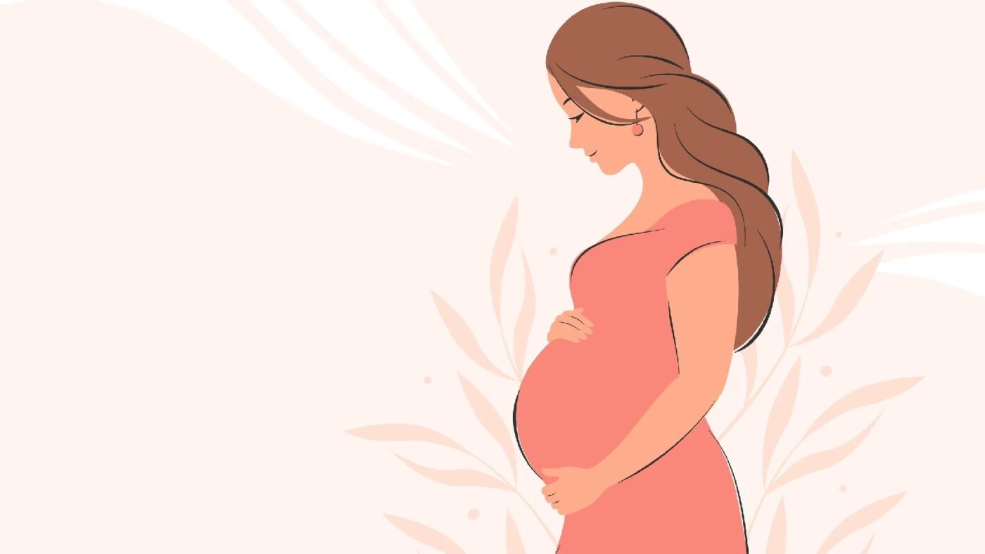
Pregnancy brings many wonderful changes, but it can also bring some discomforts, especially as the body adapts to the growing baby. Common complaints include swelling, water retention, and aching joints. An effective and non-invasive treatment for these issues is lymphatic drainage massage (MLD), which can offer relief, promote wellness, and help pregnant individuals feel their best. In this blog, we’ll explore the safety, benefits, and potential contraindications of lymphatic drainage massage during pregnancy, as well as how this therapy can be customised to suit each individual’s needs. The Benefits of Lymphatic Drainage Massage During Pregnancy Lymphatic drainage massage focus in boosting the work of the lymphatic system. By increasing the functionality of this body system, we promote the flow of lymph liquids and help the body to move unwanted substances, as well as reduce fluid retention. This type of massage has been shown to have specific benefits for pregnant individuals (Cataldo Oportus et al., 2013), particularly when it comes to managing swelling and improving overall circulation. The benefits of include: Reduction of Swelling (Oedema): One of the most common pregnancy-related discomforts is swelling in the feet, ankles, and legs. Lymphatic drainage helps to enhance the flow of lymph, which can reduce fluid buildup in the tissues and alleviate discomfort caused by oedema. Relief from Pregnancy-Related Back Pain: Lymphatic drainage massage can reduce tension in the back and promote better posture, helping to relieve pain caused by the increased weight and pressure on the spine during pregnancy. In this case, the treatment is delivered in a side position. Improved Circulation: Pregnancy can lead to changes in blood flow and circulation. Lymphatic drainage promotes circulation and can improve oxygen and nutrient delivery to both the mother and baby. Mental and Emotional Wellbeing: Lymphatic drainage massage is extremely relaxing, and this positive side effect of the treatment can help you in having a better sleep which obviously can overcome stress and body ache. Per standard and comfort, at Melbourne Massage and Treatment, I tend to deliver lymphatic drainage massage in a seated position for pregnant women. This is possible thanks to the reclined hydraulic table, which can be converted from a flat table into a comfortable and big chair. How Compression Stockings Can Help Reduce Swelling Another effective way to manage swelling during pregnancy is the use of compression stockings. I recommend and provide to my patient stockings from Sigvaris, which is one of the world’s leading stocking manufacturers. The idea of using stocking, is to maintain pressure and liquid moving along the day, while sitting or walking, or doing your daily activities. How the stocking works is by adding a constant compression, which still leave you with a comfortable feeling and does help in pushing the liquid towards the upper body, reducing the chances of fluid accumulation in the lower limbs. For pregnancy, the grade recommended is grade 2, which is a clinical grade stocking. Consider that grade 3 is what is recommended for presentation like Lymphoedema. In terms of measurements, I can easily take mesuraments on site for the best fitting compression, and have the leggings with in 2 to 3 business days. For any enquire about the product or the services please do not hesistate to contact me now. What to Consider When Delivering a Massage During Pregnancy Now we are going to talk about what to consider when we deliver a massage to a pregnant patient, and as you will read, often light pressure is a must in this type of treatment, which gives lymphatic drainage massage an advantage compared to traditional relaxation pregnancy massage. So, there are certain areas of the body that we tend to avoid working on during a massage treatment for pregnancy, or where we may work on, but with a really light pressure and a full verbal or sometimes written consent. Here is a brief summary: 1. Abdominal Area: Deep or direct pressure on the abdominal area is a big NO. Instead, we can do gentle, light strokes if the patient is comfortable with it and they request it. Explicit consent here is a must. 2. Lower Back (Spine and Sacral Area): This is another big NO for heavy pressure. The reason lies behind the fact that strong pressure can lead to strain of the ligaments that hold the joints together, which, as the pregnancy progresses to a later stage, become more and more relaxed, allowing the birth to happen. So again, gentle massage techniques such as effleurage (long, sweeping strokes) or light kneading can be beneficial, but light touch. 3. Legs: Inner thighs another area where we avoid strong pressure, due to the presence of pressure points that could potentially stimulate uterine contractions if over-stimulated (e.g., peroneal or acupressure points). 4. Ankles and Feet: Ankles and feet, as there are acupressure points (like those related to the reproductive system) that may lead to unwanted effects if stimulated too forcefully. That said, a gentle foot massage can help relieve swelling and discomfort. 5. Hand and wrist The area of your hand, between thumb and index, and the pulsing point on the wrist, are other areas where massage is not recommended as it can cause contractions. Always remember that this is a brief summary, and everyone can react differently to the work received in that area. Therefore, there is no need to be scared of making contact with other people, and you should feel comfortable with your body. Trimesters and Positioning The position to be in during a massage, including lymphatic drainage massage, also depends on the trimester in which you are. First Trimester (0–12 Weeks): Lying on the back or side is generally fine during the first trimester, as long as the client is comfortable. A reclining position with support (e.g., pillows or bolsters) is often preferred for comfort. Lying prone (on the stomach) can be done during the first trimester if the client feels comfortable. However, lying flat on the stomach will become uncomfortable and impractical as pregnancy progresses. Second and Third […]

May
Muscle tension headache and migraine are two different types of presentation that have in common a pain, which can also be debilitating, in the head area. Back in 2019, in Australia, 3 million people were estimated to suffer from migraine (Wijeratne et al., 2023), where, define how many people are suffering from muscular tension head-ache is a bit more tricky, as is not a presentation that can be easily tracked, due to self managed protocols, and other miss data counting. That said, they have different origins, symptoms, and treatment options. In this blog post, we will explore the key differences between muscle tension headaches and migraines, helping you understand how to identify and manage them. What Are Muscle Tension Headaches? Muscle tension headaches, or tension-type headaches, are the most common. This type of headache originates from cervical or facial muscle tensions, which recreates a pattern of pain on the head of facial area. As with all muscles, but even joints, the pain that we can experience can be local or in an area around the tense spot. These headaches are often linked to stress, lack of good posture, anxiety, and even sleep disturbances. They can be chronic or occasional, but compared to migraine, they lack neurological symptoms. Symptoms of Muscle Tension Headaches: Dull, aching pain or pressure around the head, especially in the forehead, temples, and back of the head. A sensation of tightness or “band-like” pressure around the head. Mild to moderate intensity (usually not as severe as a migraine). Pain can last from 30 minutes to several hours, sometimes even days. Tenderness or tightness in the neck, shoulders, and scalp. Causes of Muscle Tension Headaches: Stress: Emotional and mental stress is one of the primary causes of muscle tension in the neck and scalp muscles. Lack of good posture: Sitting or standing with poor posture and lack of strength in the musculoskeletal system, especially for long work, can strain muscles and trigger headaches. Sleep issues: Sleep deprivation or poor-quality sleep can exacerbate muscle tension and lead to headaches. The body recovers from the fatigue of the day before during sleep, especially in the early morning hours. Sleep deprivation would increase the chance of a headache. Dehydration: Not drinking enough water can lead to tension and headache symptoms. The body withdraws water from the brain to keep the organ functioning, causing physical brain shrinkage, which leads to headaches. Recent studies have indicated that chronic tension-type headaches (CTTH) are often exacerbated by environmental stressors, and poor posture in daily activities can cause muscle imbalance and contribute to the frequency of these headaches (Bendtsen et al., 2018; Grazzi et al., 2016). Treatment Options: Pain relief: Over-the-counter pain relievers, like ibuprofen or acetaminophen, can help ease the discomfort. Heat pack: Applying a warm compress to the neck and shoulders can help relax tense muscles. Keep always in mind that heat application should be limited to 10-15 minutes, once or twice a day. Massage: Gentle massage of the neck and shoulder muscles can reduce tightness and alleviate headache symptoms. Stress management: Practising relaxation techniques such as deep breathing, thai yoga, and meditation can reduce stress and prevent muscle tension headaches. Strengthen muscles: Strengthening the muscles around your cervical and shoulder area can help reduce the chance of suffering a headache by reducing the inflammatory response that the muscle would activate due to a lack of strength. What Are Migraines? As I mentioned above, the significant difference between headaches and migraines is due to neurological symptoms, a unique characteristic of migraines. Migraines are neurological events that involve complex brain activity. They are characterised by intense, throbbing pain, usually on one side of the head. They are often accompanied by other symptoms such as nausea, vomiting, and sensitivity to light and sound. Migraines are more debilitating than muscle tension headaches and can last a few hours to several days. The intensity of the headache doesn’t have to be severe. Symptoms of Migraines: Although many people experience nausea, vomiting, and light sensitivity, migraine symptoms can vary, with some individuals experiencing dizziness or visual disturbances without significant head pain. Throbbing or pulsing pain, usually on one side of the head. Nausea and vomiting. Sensitivity to light, sound, and sometimes smells (aura). Visual disturbances such as flashing lights or blind spots (this is known as an aura, which can occur before or during the headache). Dizziness or feeling lightheaded. Migraines are understood to be primarily driven by neurovascular changes and neuronal hyperexcitability (Feng et al., 2021). A review by Wagner et al. (2021) found that the pathophysiology of migraines involves alterations in neurotransmitter systems, notably serotonin and CGRP (calcitonin gene-related peptide), which contribute to the vasodilation and pain signaling pathways. Causes of Migraines: Genetics: Migraines tend to run in families, suggesting a genetic component. Hormonal changes: For many women, changes in estrogen levels, such as during menstruation, pregnancy, or menopause, can trigger migraines. Environmental triggers: Bright lights, strong smells, certain foods (like chocolate, cheese, or caffeine), weather changes, lack of sleep, and allergies that cause sinus issues are common migraine triggers. Neurological factors: Migraines may involve changes in the brain’s nerve pathways, chemicals, and blood vessels, which cause inflammation and pain. Treatment Options for Migraines: Prescription medications: Triptans and anti-nausea medications are commonly prescribed to treat the acute pain of migraines. Preventive medications: For frequent migraine sufferers, medications such as beta-blockers, antidepressants, or anti-seizure drugs may be prescribed to reduce the frequency and severity of attacks. Lifestyle changes: Regular sleep, a healthy diet, and consistent exercise can help reduce the frequency of migraines. Cognitive-behavioural therapy (CBT): Managing stress through therapy can help alleviate migraine triggers. Alternative therapies: Acupuncture, biofeedback, and massage therapy are sometimes used as complementary treatments for migraine management. Recent studies support preventive treatments for chronic migraines, such as CGRP antagonists (Kundera et al., 2020) and neuromodulation techniques like transcranial magnetic stimulation (Lefaucheur et al., 2017). Key Differences Between Muscle Tension Headaches and Migraines Although muscle tension headaches and migraines involve head pain, they differ […]

Apr
Menopause is a natural phase in every woman’s life, but it comes with a variety of physical, emotional, and mental challenges. As hormone levels shift, particularly estrogen, progesterone, and testosterone, many women experience symptoms like hot flashes, mood swings, sleep disturbances, and weight gain. Fortunately, for managing menopause symptoms, various lifestyle changes can be put in place, such as exercise, a balanced diet, improved sleep hygiene, and stress management techniques. In this blog, we will explore how adopting a healthier lifestyle can significantly improve your menopausal experience. Exercise and Physical Activity For Managing Menopause Symptoms Exercise is again the best recommendation for health improvement that can be offered here. Of course, implementing exercises alone without following any other changes or advice (where needed) is not going to do the trick. But let’s start from here. Exercises can improve overall health and have specific benefits that help ease common issues like weight gain, mood swings, and hot flashes. But not only that. Indeed, exercises, and in particular strength training, are positive stress, which allows the body to regenerate and ensure the slowing down of bone and muscle mass, which is one of the main issues that a woman going through menopause is going to face. Here is a list of exercise routines that you could focus on. But keep in mind that if you really want to choose, I would strongly suggest Strength Training. Aerobic Exercise:Walking, swimming, cycling, or jogging would help in improving circulation, promote heart health, and relieve stress (for the last one, especially if done in open-air environment). Regular aerobic exercise has also been shown to reduce the frequency and intensity of hot flashes. Plus, it aids in weight management, especially during menopause when metabolism slows. Strength Training:Engaging in strength training exercises, such as weightlifting or resistance band workouts, helps preserve muscle mass and improve bone density, which decreases as estrogen levels decline. This is essential for reducing the risk of osteoporosis and maintaining strong bone and muscle mass. Yoga or Pilates:Both yoga and Pilates are great for improving flexibility, balance, and muscle strength while reducing stress and anxiety. These low-impact exercises help maintain your physical health and mental well-being, both of which can be affected by hormonal fluctuations during menopause. Tai Chi or Qigong:These ancient practices involve slow, deliberate movements and deep breathing. They are excellent for enhancing balance, reducing stress, and promoting relaxation—especially helpful for managing mood swings and anxiety. Eating an Anti-Inflammatory Diet A nutrient-rich diet can significantly help in managing menopause symptoms. By incorporating specific foods and avoiding certain triggers, you can reduce inflammation, balance hormones, and support your body’s needs during this transition. Phytoestrogens:Phytoestrogens are plant-based compounds that mimic estrogen in the body and can help alleviate symptoms like hot flashes and night sweats. Foods rich in phytoestrogens include soy products (tofu, tempeh, edamame), flaxseeds, lentils, chickpeas, and whole grains. Calcium and Vitamin D:As estrogen levels decline, the risk of bone loss and osteoporosis increases. To support bone health, incorporate calcium-rich foods such as leafy greens (kale, broccoli, sesame seeds), fortified plant-based milks, and dairy products. Vitamin D is crucial for calcium absorption, so get it from sun exposure or foods like fatty fish (salmon, mackerel) and fortified foods. Vitamin D is cumulative, so during the longer days of the year, ensure to spend some extra time in the sun. But of course, do so during the safest hours and not at UV light pick time. Regarding Vitamin D and Calcium supplements, there is a strong debate about whether they are good or what potential side effects they have, so you’d better talk to your GP about the specifications. Healthy Fats:Omega-3 fatty acids found in fatty fish, flaxseeds, chia seeds, and walnuts have anti-inflammatory properties that can help reduce joint pain and inflammation during menopause. These healthy fats also support heart health, which is increasingly important as estrogen levels drop. Whole Grains and Fibre:Fibre helps stabilise blood sugar levels and improves digestion, which can be helpful as metabolism slows. Incorporate fiber-rich foods like whole grains (brown rice, oats, rye), fruits, vegetables, and legumes to support digestive health and reduce bloating. In the case of beans and grain, ensure that they are soaked when needed to reduce the bloat side effect. Limit Sugary and Processed Foods:Foods high in sugar and processed carbs can cause blood sugar spikes and crashes, leading to irritability and fatigue. This is where having a variety of fresh food is a key component. So yes, no one wants you to overstress about what you eat or not, but, generally speaking, if you have never looked into a balanced anti-inflammatory diet, it is time to do so. Reducing Alcohol and Inflammatory Foods Both alcohol and inflammatory foods can exacerbate menopause symptoms, so limiting or avoiding them can provide significant relief. Limit Alcohol:While alcohol might seem like a way to unwind, it can actually trigger hot flashes, disrupt sleep, and contribute to mood swings. Moderation is key—try limiting your alcohol intake to no more than one drink per day, and if possible, reduce it further to see if it improves your symptoms. Alcohol is a substance that the body does not recognise, and it has quite a hard time breaking it down. Avoid Inflammatory Foods:Highly processed foods, refined sugars, and trans fats can increase inflammation in the body and worsen menopause symptoms like joint pain, fatigue, and mood swings. Instead, focus on anti-inflammatory foods such as berries, leafy greens, fatty fish, and nuts to support your body during this phase. Inflammatory foods are all those that contain Omega-6 fatty acids, which are long-chain fatty acids, that would get collected in your Lymphatic System at first, and attacked by the macrophages (white cells), inciting an inflammatory response. Caffeine:Excessive caffeine can disrupt sleep and worsen hot flashes. If you find that caffeine aggravates your symptoms, consider reducing your intake or cutting back on coffee and other caffeinated beverages, especially in the afternoon or evening. Here is a great podcast from ZOE podcast about […]
Mar
Musculoskeletal pain can be complex, and orthopedic tests and hands-on treatment, sometimes, can be a limited tool to individualise what is happening with the body’s internal structure. Indeed, there are times when a deeper look is required to ensure we are on the right path. This is where body scans imaging comes into play to identify presentations like tendinopathy, bursitis, ligament tear or other underlying conditions. The Role of Body’s Scan in Diagnosing Pathology Body scans include a series of imaging technologies, such as ultrasound, x-ray, MRI, to name a few. Ultrasound is a highly effective imaging tool used to assess soft tissue structures in real-time. Unlike X-rays, which primarily show bone, ultrasound provides detailed images of muscles, tendons, bursae, and ligaments. This makes it an excellent tool for diagnosing conditions such as: Tendinopathy – A chronic condition involving tendon degeneration due to overuse or injury. Bursitis – Inflammation of the bursae, the small fluid-filled sacs that reduce friction between tissues. Those tissue types are found along different body joints, like the shoulder and the hip. Ligament Tears – Partial or complete tears of ligaments, often occurring after trauma or excessive stress. Baker’s cyst – is a fluid-filled swelling that forms behind the knee, often resulting from knee joint conditions like arthritis or meniscal tears, causing discomfort and limited mobility. When we are suspicious of one of those presentations, due to positive results obtained by orthopedic test and medical history, including mechanism of injury, we attempt a recovery process, based on the type of injury, symptoms, and other relevant information. Along this recovery process, we may start with isometric exercises. If, with the first 6 weeks, and a series of sessions, 3 to 4 sessions with this time frame, we still don’t see a major recovery, then we may want to get extra investigation ongoing via an ultrasound scan, which can clarify the underlying pathology. It allows us to confirm or rule out certain conditions, ensuring that treatment strategies are aligned with the actual tissue damage (if any is present). On the other hand, based always on the individual case, we could also require X-rays, which are often more helpful in diagnosing conditions related to the bones, such as arthritis or fractures, as they provide a clear view of bone structure and joint spaces. MRI is a scan that is used for Brain imaging, and when the investigation needs higher details, like when looking at the spine or a joint that via ultrasound was not giving any sign of issue. Ultrasound is also comparable to MRI, as it is faster, easier to deliver, and has fewer complications. How can myotherapy treatment help recovery from what a body scans would show? As we already discussed in another blog, Myotherapy is a practice that looks into the well-being of the skeletal muscle structure. To understand what can be done about a painful presentation, we would initially take a detailed clinical history, then look into objective measurements, such as your movement and body presentation. Given the result we can obtain, we would build up a treatment plan which includes: Hands-on Treatment – Techniques such as deep tissue massage, myofascial release, and dry needling can help reduce pain and improve mobility. Exercise Prescription – Strengthening and mobility exercises help restore function and prevent future injuries. Load Management Strategies – Proper guidance on activity levels ensures tissues heal without excessive strain. That management technique would then be combined and adjusted around the scan’s results. Here are a few examples: Bursitis: If a bursitis is confirmed, medications may be given to reduce the inflammation of the bursa, for that, we concentrate on MLD treatment to further reduce the inflammation and exercises to build strength on the structure that needs support. Ligament tear: When talking of ligament tear, the healing time can dilagate to months if not also a year, so we know now why the 6 weeks program may was not as responsive. We will keep focusing on the strength of the muscle that surrounds the specific joint, and use hands-on treatment to boost blood to the area affected. Arthritis: Medication or dietary change may be put in consideration for pain management and inflammatory reduction. Also in this case, MLD can be used to manage the pain response, and exercises for mantain movement in the affected joint/s. When Should You Consider an Ultrasound or other body scans? If you experience ongoing pain, swelling, or restricted movement that is not improving with therapy, an ultrasound or other scan helps identify the cause. This can prevent prolonged discomfort and allow for a more targeted treatment approach. At Melbourne Massage and Treatment, in Fitzroy North, we aim to provide the most effective care possible. If you’re dealing with persistent musculoskeletal pain, book a consultation with Giovanni today. Together, we’ll determine the best action to get you back to optimal function. Frequently Asked Questions (FAQs) About Musculoskeletal Pain and Body Scans Imaging 1. What are body scans, and how do they help diagnose musculoskeletal pain?Body scans include imaging technologies such as ultrasound, X-ray, and MRI. These scans help diagnose soft tissue injuries (like tendinopathy, bursitis, and ligament tears) or bone-related conditions (such as fractures or arthritis). They provide a clearer picture of what might be causing pain, inflammation, or restricted movement. 2. Why is ultrasound commonly used in diagnosing soft tissue injuries?Ultrasound is highly effective for real-time imaging of soft tissues like muscles, tendons, bursae, and ligaments. It helps diagnose conditions such as tendinopathy, bursitis, and ligament tears, providing a dynamic view of the area being studied without the need for invasive procedures. 3. When should I consider getting an ultrasound or other scans for my injury?If you’re experiencing persistent pain, swelling, or limited mobility that isn’t improving with initial therapy (such as exercises or hands-on treatment), it might be time to consider an ultrasound or other scans. These can help identify the underlying cause of your symptoms and allow for a more targeted treatment approach. 4. How do orthopedic […]

Feb
Lymphoedema and Lipoedema are chronic conditions characterized by swelling and fat accumulation, respectively, often accompanied by inflammation. Thanks to emerging research, we do not know that dietary choices, particularly the consumption of long-chain fatty acids, can influence the inflammatory processes associated with these conditions. This is why it is important to consider an Anti-Inflammatory Diet when suffering from those presentations. The Role of Long-Chain Fatty Acids in Inflammation Long-chain fatty acids are absorbed into the lymphatic system in structures called chylomicrons. Once these chylomicrons are processed, the released fatty acids can interact with macrophages—immune cells responsible for detecting and responding to pathogens. This interaction can trigger an inflammatory response, contributing to the chronic inflammation observed in both lymphoedma and Lipoedema. Why does the Lymphatic System absorb Long-Chain Fatty Acids? The lymphatic system plays a crucial role in the absorption and transport of dietary fats, particularly long-chain fatty acids. This process is essential for efficient lipid metabolism and overall energy distribution in the body. Absorption of Long-Chain Fatty Acids Long-chain fatty acids are released from dietary fats in the small intestine during digestion. These fatty acids are absorbed by the enterocytes (intestinal cells), where they are reassembled into triglycerides and packaged into lipoprotein particles known as chylomicrons. Due to their size and composition, chylomicrons are too large to enter the blood capillaries directly. Instead, they are absorbed into specialized lymphatic vessels called lacteals, located within the villi of the small intestine. This lymphatic absorption allows the efficient transport of large lipid molecules into the systemic circulation. Transport Through the Lymphatic System Once inside the lacteals, chylomicrons travel through the lymphatic system, merging into larger lymphatic vessels and eventually entering the bloodstream via the thoracic duct, which empties into the left subclavian vein near the heart. This pathway enables the gradual release of lipids into the circulation, allowing tissues to access these essential nutrients for energy production, cell membrane synthesis, and other vital functions. Benefits of an Anti-Inflammatory Diet Adopting an anti-inflammatory diet can help mitigate these effects by reducing the intake of pro-inflammatory long-chain fatty acids and emphasizing foods that support lymphatic health. Key components of such a diet include: High Fiber Intake: Consuming fruits and vegetables rich in fiber promotes the production of short-chain fatty acids in the gut, which have anti-inflammatory properties. Omega-3 Fatty Acids: Incorporating sources of omega-3s, such as fatty fish, flaxseeds, and walnuts, can reduce inflammation and edema. Anti-Inflammatory Spices: Spices like turmeric, garlic, and curry leaves possess natural anti-inflammatory effects and can be beneficial additions to the diet. Foods to Limit or Avoid for a good Anti-Inflammatory Diet To further reduce inflammation, it’s advisable to limit the consumption of: Processed Foods: Often high in trans fats and refined sugars, these can exacerbate inflammatory responses. Excessive Salt and Caffeine: High intake of salt and caffeine may contribute to fluid retention and should be moderated. Alcohol and Sweets: These can increase inflammation and are best consumed in moderation. Personalized Nutritional Guidance As a certified lymphoedema therapist trained by the Vodder Academy, I understand the importance of a holistic approach to managing lymphoedma and Lipoedema. Integrating an anti-inflammatory diet tailored to your individual needs can play a crucial role in reducing inflammation and improving overall health. For personalized advice and support, consider consulting with a healthcare professional or a registered dietitian experienced in managing these conditions. By making informed dietary choices, you can actively contribute to managing inflammation and supporting your lymphatic health. For more insights on managing lymphoedema and Lipoedema through diet, you might find this video informative. Melbourne Massage and Treatment and Lymphoedema/Lipoedema presentation Even though I am not a dietitian or nutritionist, so I can not give any direct recommendations on your diet or food intake, as a Lymphoedema therapist, I can still help you manage your presentation by offering services like Manual Lymphatic Drainage (MLD) and Combine Decongestive Therapy (CDT). So, if you need to improve your Lymphoedema or Lipoedema presentation, book your free 15-minute phone consultation now to understand how Melbourne Massage and Treatment services can benefit you. FAQ about Anti-Inflammatory Diet and Lymphoedema/Lipoedema presentation Q: How do dietary choices influence inflammation in lymphoedema and Lipoedema? A: Dietary choices play a significant role in modulating inflammation associated with lymphoedema and Lipoedema. Consuming foods high in long-chain fatty acids can lead to their absorption into the lymphatic system, where they may interact with macrophages—immune cells responsible for detecting and responding to pathogens. This interaction can trigger an inflammatory response, contributing to the chronic inflammation observed in both conditions. Adopting an anti-inflammatory diet can help mitigate these effects by reducing the intake of pro-inflammatory foods and emphasizing those that support lymphatic health. Q: What are long-chain fatty acids, and how do they affect inflammation? A: Long-chain fatty acids are a type of fat molecule commonly found in various foods, including certain oils, meats, and processed products. When consumed, these fatty acids are absorbed into the lymphatic system in structures called chylomicrons. Once processed, the released fatty acids can interact with macrophages, triggering an inflammatory response. This process can exacerbate the chronic inflammation associated with lymphoedema and Lipoedema. Q: Which foods are high in long-chain fatty acids and should be limited? A: Foods rich in long-chain fatty acids that may promote inflammation include: Certain oils Meats Processed products Limiting the intake of these foods can help reduce inflammation. Q: What are the key components of an anti-inflammatory diet that are beneficial for lymphoedema and Lipoedema? A: An anti-inflammatory diet focuses on incorporating foods that help reduce inflammation and support lymphatic health. Key components include: High Fiber Intake: Consuming fruits and vegetables rich in fiber promotes the production of short-chain fatty acids in the gut, which have anti-inflammatory properties. Omega-3 Fatty Acids: Incorporating sources of omega-3s, such as fatty fish, flaxseeds, and walnuts, can reduce inflammation and edema. Anti-Inflammatory Spices: Spices like turmeric, garlic, and curry leaves possess natural anti-inflammatory effects and can be beneficial additions to the diet. Q: Are there specific foods I should […]