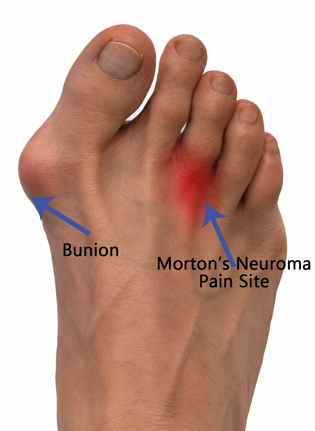
While a bunion and a Morton’s neuroma are two different foot conditions, if they are co-existing, they can sometimes influence the way the forefoot is loaded, causing delays or difficulties in the recovery process. Starting with the bunion, which medically is known as hallux valgus, changes the position and function of the big toe and first metatarsophalangeal joint. On the other hand, Morton’s neuroma is a painful condition involving one of the common plantar digital nerves, most commonly between the third and fourth toes. If you present with both conditions, treating the neuroma without considering how the rest of the foot is functioning may not always address the mechanical factors contributing to your symptoms. This is what this blog is going to analyse: Can a bunion make it harder for a Morton’s neuroma to settle? The answer is not as simple as saying that a bunion causes a neuroma, as there is not enough evidence for it. But what we know is that hallux valgus can alter how load is distributed through the forefoot, and this may be relevant when managing a sensitive or irritated interdigital nerve. What is the relationship between a bunion and a Morton’s neuroma? As mentioned above, a bunion does not automatically cause a Morton’s neuroma. That said, both conditions can be influenced by the way forces are distributed through the forefoot. Indeed, Hallux valgus can change the position of the big toe and first metatarsal, which are sitting more laterally than they should be, and alter normal walking mechanics. We know, in fact, that while walking, the big toes should be the primary driver and taker of the body weight at the toe level, and if pushed to a lateral position, it can’t take such load; therefore, the body weight is then redistributed along the phalangea box and other toes. But to be more specific, and by following what the data show, we better understand this 2025 systematic review and meta-analysis involving more than 6,000 feet found that hallux valgus was associated with reduced hallux loading when measured inside footwear. But evidence that hallux valgus consistently increases loading beneath the central metatarsals was inconclusive. And this is important because it gives a distinction between bunion and Morton’s neuroma formation, as neither is the consequence of the other. Basically, the correlation and causation effect. Indeed, foot mechanics are individual. Your symptoms can be influenced by the shape of your foot, footwear, activity, walking pattern, joint mobility, strength, and how you distribute your body weight while moving. How can a bunion affect Morton’s neuroma recovery? Now that we clarify the distinction between the correlation and causation of Morton’s neuroma from hallux valgus, we can dig more into how the bunion can affect Morton’s neuroma recovery. Morton’s neuroma is a condition that can not be recovered by only exercises or some other external interventions, but, to start with, it needs rest. By rest, we mean reducing stress on the structure involved, not no movement at all. This is why footwear modification and pressure redistribution are commonly used in Morton’s neuroma management. Therefore, the bunion may become relevant because hallux valgus can alter the way the forefoot loads during walking. For example, some people with hallux valgus develop a tendency to move the centre of pressure away from the first metatarsophalangeal joint during late stance. Research has demonstrated this lateral shift in people with bilateral hallux valgus. As a result, if the big toe can not contribute effectively to propulsion, the body may find another way to complete the movement. So again, this can overload the neuroma site, limiting rest and slowing recovery. Think about the whole foot, not just the painful spot When dealing with a painful presentation, I always invite my client to stop focusing only on the painful spot, and start to understand that nothing work in isolation. Therefore, if someone has a neuroma, it can be tempting to focus entirely on the area between the toes. But a better approach would be asking: Why is this area becoming irritated? The answer is often made up of a combination of factors, which may include: Footwear used Training load or walking volume Forefoot structure or movement Possible pre-existing injury So that’s why treating the neuroma alone may not be enough if the mechanical contributors are still present. Can bunion exercises help reduce stress on a Morton’s neuroma? The short answer is yes, but it is not always as simple. Evidence suggests exercise can improve pain and some measures of hallux valgus, particularly in mild-to-moderate cases. However, this is different from saying that bunion exercises have been proven to treat Morton’s neuroma. So what we can therefore assume is that by improving the foot mechanics and load, we may reduce stress on the neuroma site, assisting the body’s natural recovery. Exercises may include: controlled big-toe movement exercises to improve intrinsic foot muscle control toe spreading exercises strengthening exercises for the foot controlled calf and ankle exercises balance exercises progressive weight-bearing exercises exercises that improve the way the foot controls load during walking And there is no one-size-fits-all. What we should focus on too is the ability and capacity of the individual, and then work on what they are missing out on. As an example, forcing the big toe into a position with a toe separator simply because someone has a bunion is not necessarily appropriate, and it may not create the desired change anyway. The aim is not to “push the bunion back into place”. The aim here is to improve the available movement and control of the foot and help the person tolerate load more effectively, yes, starting from the big toe, if a bunion is present, but the focus has to be also on the other toes, as well as the ankle. Can big toe mobility affect pressure on a Morton’s neuroma? For what we know, the big toe plays an important role during walking, particularly during the later part of stance. For simplicity, we expect […]
Monthly Archives: September 2026

Blog
Facet Joint Irritation and Muscle Weakness: What You Need To Know
In my experience as a Clinical Myotherapist at Melbourne Massage and Treatment Myotherapy and Lymphoedema [...]
Continue readingOct

Blog
Chronic Pain: What it is, and what can you do about it?
Pain is something that we all experience, and it can start after an injury, during [...]
Continue readingOct
Blog
Bunion and Morton’s Neuroma: Can a Bunion Affect Neuroma Recovery?
While a bunion and a Morton’s neuroma are two different foot conditions, if they are [...]
Continue readingSep
Blog
Knee Pain in Coburg
Knee pain is a common form of injury and can affect people of all ages [...]
Continue readingSep

Blog
Post-Surgery MLD Melbourne – Vodder MLD Therapist
Any form of surgery is an invasive procedure for the body to deal with, and [...]
Continue readingSep
Sep
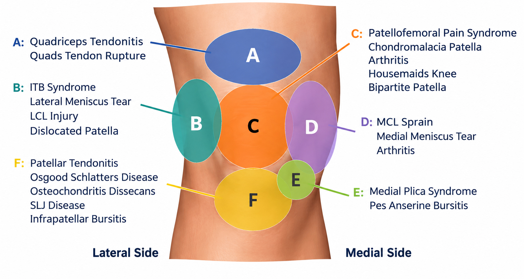
Knee pain is a common form of injury and can affect people of all ages and activity levels. Common mechanisms of action that lead to knee pain include an intense increase in load training, repetitive activities, osteoarthritis, a sudden change in direction during a sport activity, muscle or tendon tears, ligament injury or irritation around the kneecap. If you are experiencing knee pain in Coburg, the first step is understanding what may be contributing to your symptoms rather than simply treating the painful area. At Melbourne Massage and Treatment in Coburg, I use a combination of clinical assessment, hands-on treatment and exercise to help manage knee pain. Depending on your presentation, this may include Remedial Massage, Myotherapy and a progressive exercise program. What can cause knee pain? “Knee pain” is a symptom, not a diagnosis. As with any joint pain presentation, first things first, we want to rule out whether what we are looking at is a joint pain presentation or a muscle-related injury type of pain. To do so, we need an extensive understanding of the mechanism of injury, like how/when the pain started and how/when the pain increases or decreases. That information can start giving us some clues about what type of injury we may rule in or rule out Here is a list of common areas or presentations to which knee pain can refer to: Pain around or behind the kneecap Patellofemoral pain Knee osteoarthritis Patellar tendon pain Quadriceps or hamstring-related problems Pain following an increase in running, walking or gym training Knee pain following an injury Pain associated with changes in lower-limb strength or movement Stiffness and reduced knee function To give you a better understanding of how each area of the knee can correspond to a specific injury, let’s look at patellofemoral pain, which commonly becomes more noticeable during activities that load the knee, including squatting, stairs, running, jumping or prolonged sitting. But your pain, perhaps, could also be from an ACL injury, and it may present at the anterior portion of the knee, and this is why I don’t use a one-size-fits-all approach to knee pain. Unless we can pinpoint why you are in pain and what structure is responsible for it, going for a random treatment plan would not help. Knee pain assessment in Coburg During a Myotherapy appointment, I look at more than just the painful spot. The assessment can include your symptoms, medical history, previous injuries, training or work demands and the movements that reproduce your pain. Other things we may look at include: Knee range of motion Hip and ankle movement Quadriceps and hamstring function Hip strength Single-leg movements Squatting and stepping movements Running or other sport-specific activities How your symptoms respond to different loads Along with that information collection and movement pattern, I will ask you to go for, we can better understand what the structure that gives you pain is: a meniscus or a ligament, as per a quad tendon or a hamstring tendon. The purpose of this assessment is not simply to find something that looks “out of alignment”. It is to understand what may be contributing to your symptoms and determine what can be changed. How can Remedial Massage help with knee pain? Remedial Massage is an ideal hands-on treatment to reduce pain and stress on muscles that surround the knee joint, which may be contributing to your symptoms. Then, based on your individual presentation, treatment may include work around the quadriceps, hamstrings, calf muscles, gluteal muscles and other relevant areas of the lower limb. What is important to notice, on the other hand, is that massage does not directly “fix” every cause of knee pain, and it should not be considered a replacement for rehabilitation when exercise is required. Research on massage therapy for knee osteoarthritis suggests that massage may provide short-term improvements in pain, stiffness and function, although the evidence is limited and the longer-term effects are less clear. [2] This is why I see Remedial Massage as one part of a broader treatment plan when appropriate. Myotherapy for knee pain Among the services that I do offer here at my Coburg clinic, Myotherapy takes a broader clinical approach to musculoskeletal pain. Rather than simply massaging the knee or surrounding muscles, treatment can combine: Assessment → hands-on treatment → movement → exercise → gradual loading` The exact approach depends on your symptoms and goals. Hands-on treatment may be used to help manage pain, muscle sensitivity or movement restrictions. Exercise can then be introduced or progressed according to what you need to return to. For someone who has knee pain when squatting, for example, the goal may not be to avoid squatting forever. Instead, we can look at how much load you currently tolerate and gradually build your capacity. For someone who has knee pain when running, the approach may involve modifying training temporarily while building strength and tolerance before progressively returning to running. Exercise is an important part of knee pain management Exercise is one of the most important components of managing many types of knee pain, and probably any form of pain. From research to clinical experience, I support exercise therapy for knee osteoarthritis and any form of knee pain with different types of load and forms of exercise, including resistance training, cycling and other exercise approaches showing benefits for pain and function. [4] Why exercise is the way to go is because we want you to be functional and capable of loading your knee with as little trouble as possible. So, by increasing the load gradually and exposing your knee joint to positive stress as a controlled load, it works in favour of our long-term goal: you being pain-free. And of course, based on your presentation, we may need to change the type of exercises, especially in the early stage, when the inflammation is still acute, the level of sensitivity is still high, and the structure it may not be ready for any random load. Therefore, the important point is that […]
Sep
Any form of surgery is an invasive procedure for the body to deal with, and finding yourself with oedema post-surgery, pain, sensitivity, stiff scars and/or fibrosis is a natural process and consequence. Those presentations indeed are also the result of a temporary effect on the body’s tissues and the lymphatic system. This is one reason Manual Lymph Drainage (MLD) may form part of a post-operative recovery plan. But there is an important distinction that is often missed: MLD is not simply a “lymphatic massage”. Indeed, when I work with clients who specifically have undergone procedures such as liposuction, abdominoplasty, breast surgery, body contouring and other cosmetic procedures, I have to explain that MLD is performed using the Dr Vodder method, which gently stimulates the swollen areas to direct the fluid towards the nearest lymph node. And not just pressing hard or applying some oil on the skin and starting to massage it in the hope of some relaxation. Because if you are someone who is recovering from cosmetic surgery, that difference matters. What happens to the lymphatic system after cosmetic surgery? Surgery is a controlled injury to the body. As a consequence of this procedure, tissues may experience inflammation, disruption of small blood and lymphatic vessels, changes in tissue pressure and temporary impairment of normal fluid clearance. Post-operative oedema is therefore not necessarily a sign that something has gone wrong. In fact, swelling is a normal part of the body’s healing response, although the amount and duration can vary considerably between individuals and procedures. Recent research has highlighted postoperative oedema as a common consequence of aesthetic surgery and has also drawn attention to the need to better understand lymphatic complications following these procedures. That’s where, as a clinician, I don’t only think of: “How do I get rid of the swelling?” But I also look into: “What is happening to this person’s tissues and lymphatic drainage at this stage of recovery?” And that’s where clinical assessment becomes important. MLD is not about squeezing swelling away As cosmetic surgery is becoming more and more popular, and many Melburnians are heading overseas to get their surgery done, I was quite shocked the first few times I had someone call for MLD post-cosmetic surgery and asked how I would push the liquid out of their body. My response was as clear as it could be: MLD is not about squeezing liquid out of your body. This is not only a dangerous and illegal procedure here in Australia, but it is definitely not how Dr Vodder MLD is designed to work. The original Dr Vodder method uses gentle, specific skin-stretching techniques applied with appropriate pressure, direction and speed. The technique is intended to assist lymph flow and drainage of tissues. This is particularly relevant after surgery because recently operated tissues are not the same as healthy, uninjured tissue. In fact, more pressure is not necessarily better. That’s where post-operative treatment needs to respect the stage of healing and the condition of the tissues being treated. Why the treatment cannot be exactly the same for everyone No single “post-cosmetic surgery MLD protocol” fits everyone. Consider the difference between someone recovering from: liposuction of the abdomen abdominoplasty breast surgery a combination of procedures body contouring liposuction of the thighs or arms The surgical areas, incisions, tissue trauma and areas of swelling can all be different. And after a few years of experience, I can tell you, I got to work with people who did a few of those surgeries all at once, and coming up with a clinical reasoning on how to move the fluid was a good challenge. On the other hand, there is to consider that there can be variation between patients, based on their general wellbeing and health status. Two people may have the same procedure but very different presentations. One person may have relatively mild swelling and good tissue mobility. Another may have significant oedema, restricted movement, areas of tenderness or developing tissue firmness. This is why post-operative MLD should be individualised rather than treated as a routine massage sequence. What Do We Look At As a Vodder Therapist? Before commencing an MLD treatment, even with a returning client, we start with more than simply locating the swollen area. First thing first, we consider the person’s overall presentation and the stage of recovery. Depending on the situation, this may include considering: The procedure What surgery was performed? Which tissues were affected? Where are the incisions and treated areas? The stage of healing Post-operative tissues change considerably over time. A technique that is appropriate at one stage may not be appropriate at another. The distribution of swelling Swelling is not always confined to the area that was operated on. The lymphatic pathways MLD is based on understanding lymphatic anatomy and drainage pathways rather than simply rubbing towards the closest lymph node. The condition of the skin and tissues Changes in tissue texture, sensitivity, mobility and firmness can provide useful information about how recovery is progressing. The person’s symptoms and function Pain, tightness, restricted movement and discomfort may influence how treatment is approached. Compression and movement MLD is not necessarily an isolated treatment. Depending on the individual’s situation, compression, appropriate movement and other elements of recovery may also be relevant. And again, this is one reason why seeing a therapist with specific lymphatic training can be different from booking a generic “lymphatic massage”. Why the direction of MLD matters The lymphatic system is a complex network, not a simple pipe running directly from the swollen area to the nearest lymph node. Indeed, when performing MLD, we use specific manual techniques and treatment sequences to influence lymphatic drainage. Our aim as MLD therapists is to direct the fluid from the congested area, away from and across the watershed (an imaginary line that divides the body into quadrants, from which the lymphatic system vessels start), aiming for the fluid to reach the lymphatic duct (the connection between the lymphatic system and the bloodstream in the […]
Sep
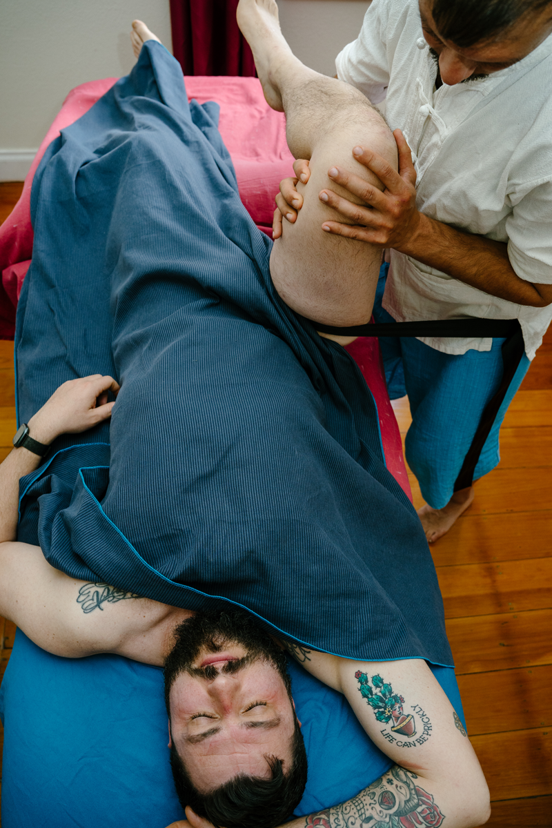
In the many presentations I get to see here at Melbourne Massage and Treatment in Coburg, hip pain is quite a common one. When we talk about hip pain, though, we refer to a variety of presentations which can be defined primarily by the site of pain in the hip area. So it can be the frontal side, including the groin, or the side of the hip, near the femoral head, or even the gluteal area, upper or lower. Hip pain, like any form of pain, can be debilitating, and as with other conditions, like back pain, it can make everyday activity more difficult and challenging, as the hip is a joint we use for basically any movement, and even while sitting or lying down can still be uncomfortable. That said, “hip pain” does not always mean that the problem is coming directly from the hip joint. Pain around the hip can involve muscles, tendons, ligaments, nerves, the sacroiliac joint (SIJ), or structures around the outside of the hip. Conditions such as femoroacetabular impingement (FAI), gluteal tendinopathy and greater trochanteric pain syndrome can also produce different patterns of hip pain. At Melbourne Massage and Treatment in Coburg, assessing where your pain is located, what movements aggravate it and how your hip and surrounding areas move can help determine what may be contributing to your symptoms. What Can Cause Hip Pain? As briefly mentioned above, there are many possible causes of hip pain and injuries. Below, we are going to go through some of the most common hip pain presentations I get to see at my Myotherapy clinic in Coburg. Ligament Tear Ligaments are soft tissues that connect bones to bones and help provide stability around joints. Even though the hip joint is most often well supported via those 3 ligaments Iliofemoral, Pubofemoral, and ischiofemoral a significant force, sudden movement, or sporting injury can damage a ligament around the hip or pelvis. When we said that most often, the hip is well supported by those 3 types of ligaments, we refer to the fact that genetically we all have different presentations, and someone who was born with hip dysplasia may present naturally with less stability at the hip joint. Moving forward, a ligament injury may cause pain, tenderness and a feeling of reduced stability. The gold standard diagnostic method for a ligament tear, lastly, would not be a basic hands-on assessment, but a magnetic resonance arthrogram (MRA). Muscle Tear The muscles surrounding the hip are among the bigger and stronger muscle groups in the body; let’s think of the quadriceps muscle group or the hamstrings. Those muscle groups, on top of being really important for overall hip functionality, are also really important for hip stability. But, as with any muscle group, a sudden increase in load, sprinting, jumping, changing direction, or an unexpected movement can result in a muscle strain or tear. Depending on the muscle involved, you may feel pain around the front, side, or back of the hip. Muscle tears can range from relatively minor strains to more significant tears, which result in severe pain, swelling, and bruising and may require a longer rehabilitation period. Hypermobility Hypermobility is a condition that involves many of my clients and is about having more laxity in the ligament tissue. However, being hypermobile does not automatically mean having an injury, but it means that more strength in the hip muscle group is needed to avoid injuries. This is because the hip ligaments, in a hypermobile presentation, are less capable of holding the femur head into the hip socket. Therefore, the goal is not necessarily to restrict movement at your hip joint, but to improve strength, control and tolerance to load. Femoroacetabular Impingement (FAI) Femoroacetabular impingement, commonly called FAI, is a condition that can affect even fairly active people, including athletes. It is characterised by extra tissue that can grow on the femoral head or on the hip socket, which may get irritated when the hip goes into flexion. Symptoms may include pain around the groin or front of the hip, particularly with certain positions involving hip flexion and rotation. In contrast to a hip ligament tear, imaging findings do not always explain someone’s symptoms on their own. A clinical assessment is needed to understand how the hip structure, movement and symptoms relate to each other. But, commonly, in ligament tears, the strengthening of muscle groups that cross the hip joint is quite important, including quads and adductors. Femoral Nerve Pain Even though femoral nerve pain is less common than sciatic nerve pain, it is still quite relevant to the topic of hip pain. A common cause of femoral nerve pain is Meralgia Paresthetica, a nerve entrapment at the inguinal ligament that can cause numbness and pain at the anterior portion of the thigh. Furthermore, femoral nerve pain can also result from back injury or nerve entrapment at the spine level, specifically L2, L3, and L4. To differentiate which pain comes from where, a tailored assessment is needed, including a clear understanding of the mechanism of injury and changes in body mass (especially if it happens in a short period of time) and, where needed, a scan can also be helpful to rule out either one or the other condition. Sacroiliac Joint (SIJ) Discomfort The sacroiliac joints are a complex structure that connect the pelvis with the sacrum and are located near the back of the pelvis. SIJ pain can present with different levels of discomfort and in different locations, including the sacral area, upper gluteal area, or, if nerve entrapment is present, pain can refer all the way down the shin via the sciatic nerve. Because symptoms from the lower back, pelvis and hip can overlap, identifying the exact source of pain can be difficult without an appropriate assessment. Gluteus Medius Bursitis / Greater Trochanteric Pain Syndrome When pain is present on the outside of the hip, it can be described as “hip bursitis” or “Greater Trochanteric Pain Syndrome”. A bursa is […]